User login
CAR T-cell therapy granted orphan designation

Photo courtesy of NIAID
The US Food and Drug Administration (FDA) has granted orphan drug designation for a CD4-directed chimeric antigen receptor (CD4CAR) T-cell therapy to treat peripheral T-cell lymphoma (PTCL).
The CD4CAR therapy, also known as ICG122, consists of properly matched allogeneic T cells engineered to express an anti-CD4 single-chain variable fragment antibody domain.
ICG122 is being developed by iCell Gene Therapeutics.
The company is planning a phase 1 trial of ICG122 in cooperation with the National Institutes of Health, Indiana Clinical and Translational Sciences Institute, Stony Brook Hospital, and the James Graham Brown Cancer Center at University of Louisville.
“CD4CAR could significantly enhance currently available treatment options for [PTCL] patients,” said Yupo Ma, MD, PhD, a professor at Stony Brook University and chairman and chief scientific officer at iCell Gene Therapeutics.
“The orphan drug designation is an important achievement as we advance our development plans for this promising treatment in T-cell hematologic cancers.”
The FDA grants orphan designation to drugs and biologics intended to treat, diagnose, or prevent diseases/disorders that affect fewer than 200,000 people in the US.
The designation provides incentives for sponsors to develop products for rare diseases. This may include tax credits toward the cost of clinical trials, prescription drug user fee waivers, and 7 years of market exclusivity if the drug is approved. ![]()
Immunotherapy conditioning proves successful in mice

Photo by Aaron Logan
Research in mice suggests it’s feasible to use an immunotherapy conditioning regimen rather than radiation or chemotherapy prior to hematopoietic stem cell transplant (HSCT).
Investigators found that combining an antibody against the HSC receptor c-Kit with a CD47-blocking therapy could eliminate host HSCs and allow for successful engraftment of donor HSCs in immunocompetent recipient mice.
Adding T-cell-depleting antibodies to the mix allowed for robust HSC engraftment in a clinically relevant model of allogeneic HSCT.
Irving Weissman, MD, of Stanford University School of Medicine in California, and his colleagues conducted this research and reported the results in Science Translational Medicine.
The researchers first found that ACK2, an antibody against c-Kit, successfully depleted HSCs in immune-deficient mice.
“However, this antibody alone would not be effective in immune-competent recipients, who represent a majority of potential bone marrow transplant recipients,” said study author Akanksha Chhabra, PhD, of Stanford University School of Medicine.
So the researchers sought to enhance the effectiveness of ACK2 by combining it with antibodies or biologic agents that block CD47. They found that blocking CD47—particularly with an antagonist known as CV1mb—liberated macrophages to engulf target cells.
In this way, the immune system effectively depleted host HSCs in the immunocompetent mice, clearing the way for donor HSCs to take up residence in the bone marrow.
Finally, the researchers set out to determine whether conditioning with an anti-c-Kit antibody and CD47-blocking therapy could be extended to a clinically relevant model of allogeneic HSCT, in which the donor and recipient are matched through human leukocyte antigen alleles but mismatched at minor histocompatibility complex (mHC) antigens.
So the team conditioned mice with either ACK2 and CV1mb or ACK2 and the anti-CD47 antibody MIAP410. And they achieved immune ablation with T-cell-depleting antibodies—GK1.5 (anti-CD4) and YTS169.4 (anti-CD8). The mice then received mHC-mismatched HSCs.
The researchers found that either conditioning regimen, when combined with a T-cell-depleting regimen, resulted in substantial granulocyte, B-cell, T-cell, and NK-cell chimerism, as well as HSC engraftment in the bone marrow.
The success of these techniques in mice raises the researchers’ hopes that similar techniques will succeed in humans.
“If it works in humans like it did in mice, we would expect that the risk of death from blood stem cell transplant would drop from 20% to effectively 0,” said study author Judith Shizuru, MD, PhD, of Stanford University School of Medicine.
“If and when this is accomplished, it will be a whole new era in disease treatment and regenerative medicine,” Dr Weissman said. ![]()
FDA authorizes use of Zika assay

Photo by Juan D. Alfonso
The US Food and Drug Administration (FDA) has granted emergency use authorization (EUA) for the xMAP® MultiFLEX™ Zika RNA Assay.
This multiplex nucleic acid test is designed to detect Zika virus RNA in blood serum, plasma, or urine (collected alongside a patient-matched serum or plasma specimen).
The xMAP® MultiFLEX™ Zika RNA Assay is available for purchase by laboratories that are certified under the Clinical Laboratory Improvement Amendments of 1988 (CLIA) to perform high complexity tests.
The assay uses the Luminex® 100/200™ analyzer, MAGPIX® system, or other authorized instruments to simultaneously test for 6 genetic targets of the Zika virus.
The xMAP® MultiFLEX™ Zika RNA Assay was designed by GenArraytion, Inc. and is marketed by Luminex Corporation.
For more information on the test, see the fact sheet for healthcare providers on the Luminex website.
About the EUA
The EUA does not mean the xMAP® MultiFLEX™ Zika RNA Assay is FDA cleared or approved.
An EUA allows for the use of unapproved medical products or unapproved uses of approved medical products in an emergency.
The products must be used to diagnose, treat, or prevent serious or life-threatening conditions caused by chemical, biological, radiological, or nuclear threat agents, when there are no adequate alternatives.
This means the xMAP® MultiFLEX™ Zika RNA Assay is only authorized as long as circumstances exist to justify the authorization of the emergency use of in vitro diagnostics for the detection of Zika virus, unless the authorization is terminated or revoked sooner. ![]()
FDA approves drug for prevention of CINV

Photo by Rhoda Baer
The US Food and Drug Administration (FDA) has approved granisetron extended-release injection (Sustol®) for the prevention of chemotherapy-induced nausea and vomiting (CINV) in adults.
Extended-release granisetron is a serotonin-3 (5-HT3) receptor antagonist that utilizes Biochronomer® polymer-based drug delivery technology to maintain therapeutic levels of granisetron for at least 5 days, covering both the acute and delayed phases of CINV.
The product is intended for use in combination with other anti-emetics to prevent acute and delayed nausea and vomiting associated with initial and repeat courses of moderately emetogenic chemotherapy (MEC) or anthracycline and cyclophosphamide (AC) combination chemotherapy regimens.
“Despite advances in the management of CINV, up to half of patients receiving chemotherapy can still experience CINV, with delayed CINV being particularly challenging to control,” said Ralph V. Boccia, MD, of the Center for Cancer and Blood Disorders in Bethesda, Maryland.
“In our experience, other 5-HT3 receptor antagonists, including palonosetron, are generally effective for 48 hours or less. Sustol, due to its extended-release profile, represents a novel option that can protect patients from CINV for a full 5 days.”
Extended-release granisetron (formerly known as APF530) is a product of Heron Therapeutics, Inc. The US commercial launch of the drug is planned for the fourth quarter of 2016.
Phase 3 trials
The global phase 3 development program of extended-release granisetron consisted of 2 large, guideline-based clinical trials of more than 2000 cancer patients.
In one trial, researchers compared extended-release granisetron to palonosetron for the prevention of acute and delayed CINV after MEC or highly emetogenic chemotherapy (HEC).
Results suggested extended-release granisetron was non-inferior to palonosetron. The most common adverse events observed in patients receiving granisetron were injection-site reactions and constipation.
In another trial, researchers compared extended-release granisetron to ondansetron for control of delayed CINV after HEC. Patients received extended-release granisetron, dexamethasone, and fosaprepitant or ondansetron, dexamethasone, and fosaprepitant.
A higher percentage of patients in the granisetron arm had delayed-phase complete response. The incidence of treatment-emergent adverse events was similar between the treatment arms.
“The Sustol clinical trial populations and results are highly representative of cancer patients in our real-world clinical practice,” said Jeffrey Vacirca, MD, of North Shore Hematology Oncology Associates in East Setauket, New York.
“Use of MEC regimens is widespread, and AC-based regimens are among the most commonly prescribed highly emetogenic chemotherapy regimens. The most significant challenge for my breast cancer patients receiving AC is chemotherapy-induced nausea and vomiting. Sustol represents a better option to manage this devastating side effect of therapy.”
For more details on the drug, access the full prescribing information at www.SUSTOL.com. ![]()
How procoagulant platelets develop
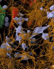
Image by Andre E.X. Brown
Researchers say they have determined how procoagulant platelets develop.
One of the mysteries in the field of thrombosis and hemostasis is how platelets are divided into two kinds when activated—“ordinary” platelets capable of aggregation and “super-activated,” procoagulant platelets.
The new study suggests that, to become super-activated, platelets must die. And the platelets need mitochondria to commit suicide.
Researchers were able to show how this programmed death—mitochondrial necrosis—follows a chain of events that lead to the platelets’ transition to a super-activated state.
“It was not clear before how a platelet makes the decision of what type to become,” said study author Mikhail Panteleev, of Lomonosov Moscow State University in Russia.
“We have deciphered the sequence of events: how the signal goes within the platelet and how the cell decides to die.”
Panteleev and his colleagues described these events in the Journal of Thrombosis and Haemostasis.
The team noted that platelets have many activators, but the chief among them are collagen, ADP, and thrombin.
Platelets detect different concentrations of an activator and respond with a varying frequency of calcium impulses in the cytoplasm.
The platelets’ mitochondria absorb and store the calcium, and when its concentration exceeds the critical level, the process of mitochondrial necrosis starts.
Calcium and reactive oxygen species are released from mitochondria, ATPases begin to destroy ATP instead of synthesizing it, the cell cytoskeleton collapses, and the platelets greatly increase in size.
As a result, at the outer membrane of the enlarged platelets, a lipid called phosphatidylserine appears, which is responsible for rapid blood clotting. And all this happens in seconds. ![]()
Stable INRs uncommon with long-term warfarin, study suggests

Results of a large study suggest most patients taking warfarin long-term do not maintain stable international normalized ratio (INR) values.
Researchers analyzed data from more than 3700 patients and found that 74% did not have stable INRs during the first 6 months of analysis.
Of the patients who did have stable INRs during this period, only 34% maintained stable INRs over the subsequent year.
Sean D. Pokorney, MD, of Duke University Medical Center in Durham, North Carolina, and his colleagues reported these findings in JAMA.
The researchers analyzed data from a prospective registry of patients with atrial fibrillation treated at 176 clinics. The patients were enrolled from June 2010 through August 2011 and followed for 3 years through November 2014.
Patients receiving warfarin at study entry with 3 or more INR values in the first 6 months and 6 or more in the subsequent year were included.
Of 10,132 registry patients, 6383 were not taking warfarin or had insufficient INR values and were excluded.
So there were 3749 eligible patients taking warfarin. Their average age was 75. Forty-three percent of patients were female, and 91% self-identified as white.
The patients’ median time from their first warfarin prescription to baseline was 3.9 years (range, 1.5-7.5 years). Thirty-seven percent of patients were also taking aspirin, and 5% were taking clopidogrel.
Results
INR stability was defined as 80% or more INRs in the therapeutic range (2.0-3.0).
Twenty-six percent of the patients met this definition during the first 6 months, and 34% of these patients maintained stable INRs over the subsequent year.
Ten percent of all patients had 100% of their INR values in the therapeutic range during the first 6 months. Of these patients, 37% met the definition of stability over the subsequent year.
Of the patients who had 80% or more of their INR values in the therapeutic range at baseline, 36% had 1 or more well-out-of-range INRs in the following year.
Of the patients with 100% of their baseline INR values in the therapeutic range, 33% had 1 or more well-out-of-range INRs in the subsequent year.
The researchers said these results suggest warfarin stability is difficult to predict, and this study challenges the notion that patients who have done well taking warfarin should continue taking warfarin. ![]()
Device could be used to monitor antiplatelet therapy
Photo courtesy of the
Wyss Institute at Harvard
A novel microfluidic device can be used to monitor thrombus formation and platelet function in patients receiving antiplatelet therapy, according to research published in Biomedical Microdevices.
The device is designed to mimic cellular and vascular flow conditions inside the human body.
Investigators say it demonstrates how endothelial cells contribute to hemostasis, even though it does not contain living endothelial cells.
The device contains microfluidic channels lined with chemically fixed human endothelial cells.
“It’s a bioinspired device that contains the endothelial function of a diseased patient without having actual living cells, and this greatly increases the robustness of the device,” explained study author Abhishek Jain, PhD, of Texas A&M University in College Station, Texas.
D Jain and his colleagues said their device retains the ability to modulate hemostasis under continuous flow in vitro, even after a few days of storage.
And the team successfully used the device to measure thrombus formation and platelet function in small amounts of whole blood from patients receiving antiplatelet therapy.
“Abnormal blood coagulation and platelet activation are major medical problems, and the ways we study them now are overly simplified,” said study author Donald Ingber, MD, PhD, of the Wyss Institute for Biologically Inspired Engineering at Harvard University in Boston, Massachusetts.
“Clinicians currently do not have tools to monitor hemostasis that take into account physiologically important interactions between endothelial cells and flowing blood.”
In a previous study, Dr Ingber and his colleagues showed that recreating the physicality and blood flow of vasculature within microfluidic channels allowed them to predict precise times that blood might clot, with potential applications in real-time monitoring of patients receiving anticoagulants.
The group’s new device adds another layer of complexity by embedding the functionality of the vascular endothelium within a tool that might be manufactured, stored, and shipped for clinical use.
“This is one of the first examples of how a microfluidic cell culture system could have added value in clinical diagnostics,” said study author Andries van der Meer, PhD, of University of Twente in Enschede, Netherlands.
“Using chemically fixed tissue that is no longer alive offers a clear, low-risk path toward further testing and product development.” ![]()
Photo courtesy of the
Wyss Institute at Harvard
A novel microfluidic device can be used to monitor thrombus formation and platelet function in patients receiving antiplatelet therapy, according to research published in Biomedical Microdevices.
The device is designed to mimic cellular and vascular flow conditions inside the human body.
Investigators say it demonstrates how endothelial cells contribute to hemostasis, even though it does not contain living endothelial cells.
The device contains microfluidic channels lined with chemically fixed human endothelial cells.
“It’s a bioinspired device that contains the endothelial function of a diseased patient without having actual living cells, and this greatly increases the robustness of the device,” explained study author Abhishek Jain, PhD, of Texas A&M University in College Station, Texas.
D Jain and his colleagues said their device retains the ability to modulate hemostasis under continuous flow in vitro, even after a few days of storage.
And the team successfully used the device to measure thrombus formation and platelet function in small amounts of whole blood from patients receiving antiplatelet therapy.
“Abnormal blood coagulation and platelet activation are major medical problems, and the ways we study them now are overly simplified,” said study author Donald Ingber, MD, PhD, of the Wyss Institute for Biologically Inspired Engineering at Harvard University in Boston, Massachusetts.
“Clinicians currently do not have tools to monitor hemostasis that take into account physiologically important interactions between endothelial cells and flowing blood.”
In a previous study, Dr Ingber and his colleagues showed that recreating the physicality and blood flow of vasculature within microfluidic channels allowed them to predict precise times that blood might clot, with potential applications in real-time monitoring of patients receiving anticoagulants.
The group’s new device adds another layer of complexity by embedding the functionality of the vascular endothelium within a tool that might be manufactured, stored, and shipped for clinical use.
“This is one of the first examples of how a microfluidic cell culture system could have added value in clinical diagnostics,” said study author Andries van der Meer, PhD, of University of Twente in Enschede, Netherlands.
“Using chemically fixed tissue that is no longer alive offers a clear, low-risk path toward further testing and product development.”
Odor-baited mosquito traps can fight malaria

Photo courtesy of the CDC
Solar-powered mosquito traps incorporating human odor can reduce the incidence of malaria, according to research published in The Lancet.
Researchers introduced these traps to homes on the Kenyan island of Rusinga.
The population of malaria-carrying mosquitoes declined by 42% in homes that had the traps.
And the prevalence of malaria was 30% lower among people living in houses with a trap than among those in houses without a trap.
“The objective of the trial on Rusinga Island in Lake Victoria was to investigate whether malaria mosquitoes can be captured and destroyed using traps with a lure so that the risk of new malaria infections is minimized,” explained study author Willem Takken, PhD, of Wageningen University and Research Centre in Wageningen, Netherlands.
The trial enrolled 34,041 participants. Each individual was assigned to a cluster, which consisted of 50 or 51 geographically contiguous households. There were 81 clusters in all.
The researchers installed their solar-powered, odor-baited mosquito trapping systems (SMoTS) in the various households, cluster by cluster, until all of the clusters had the traps.
During the roll-out period—between June 3, 2013, and May 16, 2015—SMoTS were installed in 4358 households.
The density of Anopheles mosquitoes was lower in the clusters with SMoTS than those without. The adjusted estimated effectiveness of the traps was 42.2%.
The densities of Anopheles funestus and Anopheles gambiae mosquitoes were lower in clusters with SMoTS than those without. The adjusted estimated effectiveness was 69.2% (P=0.005) and 10.8% (P=0.6), respectively.
The prevalence of malaria was 29.8% lower in clusters with SMoTS than those without (P<0.0001).
About 24% of people in clusters with SMoTS were positive for Plasmodium parasites (23.7%, 1552/6550), compared to about 35% of people in clusters without SMoTS (34.5%, 2002/5795).
“Ultimately, we want to eradicate malaria completely, in an environmentally friendly and sustainable manner,” Dr Takken said.
“As we use a natural lure—namely, human odor—in our approach, there is no negative impact on the environment, and it is very improbable that the mosquitoes will become ‘resistant’ to being captured. After all, the mosquitoes need their attraction to the lure in order to be able to survive.”
Dr Takken and his colleagues believe their SMoTS may also be able to combat dengue fever and the Zika virus. Aedes aegypti is a vector for these viruses, and this mosquito is attracted to the same humanized scent that attracts malaria-carrying mosquitoes.
Drivers of cardiac complications in sickle cell anemia

with sickle cell anemia
Image courtesy of the
University of Michigan
Preclinical research has revealed malfunctioning molecular pathways associated with cardiac anomalies in sickle cell anemia (SCA) that lead to sudden death.
Researchers used a mouse model of SCA and identified a unique “restrictive cardiomyopathy” that is superimposed on the anemia-associated heart enlargement.
They also found upregulated gene expression that fuels myocardial fibrosis and electrophysiological changes.
The researchers believe these findings, published in PNAS, will aid the development of new targeted therapies to treat cardiac dysfunction in patients with SCA.
“Sickle cell anemia is associated with significant morbidity and mortality, including a high incidence of unexplained, sudden death in young adults,” said study author Punam Malik, MD, of Cincinnati Children’s Hospital Medical Center in Ohio.
“Our findings may provide a unifying cardiac pathophysiology that explains reported cardiac abnormalities and sudden death seen in humans with SCA.”
To find pathologies specific to SCA, Dr Malik and his colleagues compared mice bred to have SCA and wild-type mice with experimentally induced chronic anemia.
The mice underwent serial comprehensive cardiac analysis, including detailed cardiac imaging (MRI), electrocardiography, and microscopic cross-section analysis of heart tissues (histopathology and electron microscopy).
The researchers said that, in the SCA mice, they observed a distinctive sickle cardiomyopathy in which restrictive physiology was superimposed on chronic anemia and predisposed the mice to sudden death.
The SCA mice had progressive left atrial enlargement and diastolic dysfunction but preserved systolic function. This restrictive physiology appeared to be caused by ischemic-scattered cardiomyocyte loss, myocardial fibrosis, and cardiac remodeling.
The researchers also conducted transcriptome analysis. This revealed that SCA mice had upregulation of genes that cause increased oxidation, hypoxia, and fibrosis in heart tissues. It also showed a downregulation of genes associated with electrophysiological function.
Finally, the team observed progressive corrected QT prolongation, arrhythmias, and ischemic changes in the SCA mice shortly before a significant number of the animals experienced sudden death.
The researchers are continuing to study the molecular mechanisms and pathways that trigger myocardial fibrosis in SCA, using a variety of knockout mice.
In addition, the successful use of noninvasive cardiac imaging techniques in this study inspired the researchers to launch a clinical trial to test these early diagnostic techniques on people with SCA. Enrollment in that study is now complete.
“It is incredibly exciting to see that this work has inspired clinical trials and other research studies,” said study author Nihal Bakeer, MD, of the Indiana Hemophilia and Thrombosis Center in Indianapolis.
“Our goal has always been [to] find the underling pathobiology of cardiac complications in sickle cell anemia and help find new diagnostics and therapeutics to decrease the morbidity and rate of sudden cardiac death in young adults with SCA.”
How hydroxyurea fights sickle cell disease

Photo by Zak Hubbard
Researchers say they have uncovered hydroxyurea’s main mechanism of action in sickle cell disease (SCD).
The drug’s mechanism has been a topic of debate, with some researchers claiming hydroxyurea works by reactivating fetal hemoglobin and others saying it increases the volume of red blood cells (RBCs), thereby reducing the concentration of sickle hemoglobin.
Now, research published in PNAS suggests the latter mechanism is the dominant one.
“Our findings shine a light on the mechanism behind hydroxyurea action, which has long been debated in the scientific community,” said study author Ming Dao, PhD, of the Massachusetts Institute of Technology in Cambridge.
“It’s exciting to see that, using the latest optical imaging tools, we can now confirm which one is the dominating mechanism. Understanding the key mechanism of action will allow us to explore novel and improved therapeutic approaches for sickle cell disease.”
For this study, the researchers analyzed blood samples from patients with SCD.
The team used common-path interferometric microscopy to assess the biophysical properties (shape, surface area, and volume) and biomechanical properties (flexibility and stickiness) of RBCs.
The researchers separated RBCs into 4 groups based on their density. Normal, disc-shaped cells were the least dense, while severely sickled cells were the densest.
The team then compares samples from patients who were taking hydroxyurea and those who were not.
The RBCs of patients receiving treatment showed an improvement in all of the biophysical and biomechanical properties tested across all density levels.
Improvement in the physical properties of RBCs from patients treated with hydroxyurea correlated more with an increase in RBC volume than with levels of fetal hemoglobin.
The researchers hope these biophysical markers can be combined with biochemical and molecular-level markers to assess the severity of a patient’s disease, determine whether or not a patient will respond to hydroxyurea, and monitor the effectiveness of that treatment.
“There is a critical need for patient-specific biomarkers that can be used to assess the effectiveness of treatments for sickle cell disease,” said study author Subra Suresh, ScD, of Carnegie Mellon University in Pittsburgh, Pennsylvania.
“This study shows how techniques commonly used in engineering and physics can help us to better understand how the red blood cells in people with sickle cell disease react to treatment, which could lead to improved diagnostics and therapies.”