User login
Edoxaban appears comparable to standard therapy

ROME—The novel oral anticoagulant edoxaban is a feasible treatment option for patients with atrial fibrillation (AF) who need anticoagulation before cardioversion, according to researchers.
Results of the ENSURE-AF trial suggest edoxaban is “an effective and safe alternative” to standard therapy with warfarin and enoxaparin and may allow for prompt cardioversion with transesophageal echocardiography (TEE), said Andreas Goette, MD, of St. Vincenz-Hospital in Paderborn, Germany.
“These results may have important clinical implications for newly diagnosed, non-anticoagulated AF patients undergoing cardioversion,” Dr Goette noted.
“According to the study protocol, a newly diagnosed, non-anticoagulated AF patient was started on edoxaban, and the cardioversion procedure was scheduled as early as 2 hours following the start of treatment, when applying a TEE-guided approach.”
Dr Goette presented results from ENSURE-AF at ESC Congress 2016 (abstract 5715). The data were simultaneously published in The Lancet. The study was supported by Daiichi Sankyo.
This phase 3b study included 2199 patients with documented non-valvular AF who were scheduled for electrical cardioversion after anticoagulation therapy.
Patients were randomized to receive edoxaban (n=1095) or warfarin with enoxaparin bridging (n=1104). Edoxaban was dosed at 60 mg once daily, but the dose was reduced to 30 mg if one or more of the following factors were present: renal impairment, low body weight, or concomitant use of certain P-glycoprotein inhibitors.
Patients were stratified according to the cardioversion approach (TEE or non-TEE), a patient’s prior experience taking anticoagulants at the time of randomization (ie, anticoagulant-experienced or naïve), and edoxaban dose (60 mg or 30 mg). Patients were randomized in a 1:1 ratio to 2 treatment groups within each stratum.
The primary efficacy endpoint was the composite of stroke, systemic embolic event, myocardial infarction, and cardiovascular death at day 28.
This endpoint occurred at a comparable rate in both treatment arms—0.5% in the edoxaban arm and 1.0% in the enoxaparin/warfarin arm (odds ratio [OR]=0.46; 95% CI, 0.12–1.43).
The primary safety endpoint was the composite of major and clinically relevant non-major bleeding events at 30 days.
This endpoint also occurred at a comparable rate in both arms—1.5% in the edoxaban arm and 1.0% in the enoxaparin/warfarin arm (OR=1.48; 95% CI, 0.64–3.55).
The result for the net clinical outcome—a composite of stroke, systemic embolic event, myocardial infarction, cardiovascular mortality, and major bleeding—was 0.7% in the edoxaban arm and 1.4% in the enoxaparin/warfarin arm (OR=0.50; 95% CI, 0.19–1.25) during the overall study period.
The researchers said the ORs recorded in this trial should be viewed with caution because the trial was not adequately powered to show statistically significant differences for efficacy or safety endpoints.
Still, the team said the results suggest edoxaban can provide an alternative to standard therapy.
“At a practical level, our study results show that newly diagnosed, non-anticoagulated AF patients can start edoxaban as early as 2 hours prior to their cardioversion procedure if they have access to [TEE] or 3 weeks prior without,” Dr Goette said. ![]()
Drug granted orphan designation for GVHD
Photo by Linda Bartlett
The European Commission has granted orphan drug designation to ALXN1007 for the treatment of graft-versus-host disease (GVHD).
ALXN1007 is an anti-inflammatory monoclonal antibody targeting complement protein C5a.
The drug is currently under investigation in a phase 2 trial of patients with newly diagnosed acute GVHD of the lower gastrointestinal tract (GI-GVHD).
ALXN1007 is being developed by Alexion Pharmaceuticals, Inc.
About orphan designation
Orphan designation from the European Commission provides regulatory and financial incentives for companies to develop and market therapies that treat a life-threatening or chronically debilitating condition affecting no more than 5 in 10,000 people in the European Union, and where no satisfactory treatment is available.
Orphan designation provides a 10-year period of marketing exclusivity in the European Union if the drug receives regulatory approval. The designation also provides incentives for companies seeking protocol assistance from the European Medicines Agency during the product development phase and direct access to the centralized authorization procedure.
Phase 2 trial of ALXN1007
Results from the phase 2 trial of ALXN1007 in patients with newly diagnosed, acute GI-GVHD were presented at the 21st Congress of the European Hematology Association (abstract LB2269).
The presentation included 15 patients with biopsy-confirmed acute GI-GVHD. The patients had a median age of 60 (range, 25-69), and 60% were male.
Patients had acute myeloid leukemia/myelodysplastic syndrome (n=8), acute lymphoblastic leukemia (n=2), acute lymphocytic leukemia (n=1), acute myeloblastic leukemia (n=1), aplastic anemia (n=1), cutaneous T-cell lymphoma (n=1), or mantle cell lymphoma (n=1).
Most patients received transplants from matched, unrelated donors (n=11), 3 had matched, related donors, and 1 had a mismatched donor. Ten patients received peripheral blood grafts, 4 received cord blood, and 1 received a bone marrow transplant.
Patients had grade 1 (n=7), grade 2 (n=2), and grade 3 acute GI-GVHD (n=6).
The patients received weekly doses of ALXN1007 at 10 mg/kg, in combination with methylprednisolone at an initial dose of 2 mg/kg, through day 56.
Thirteen patients were evaluable for efficacy. One patient experienced leukemia relapse at day 18, and 1 withdrew from the study early.
The overall acute GVHD response rate was 77% (10/13), both at day 28 and day 56. The complete GI-GVHD response rate was 69% at day 28 and 77% at day 56.
At day 180, the nonrelapse mortality rate was 12.5%, and the overall survival rate was 69.2%.
All of the patients had treatment-emergent adverse events (AEs), and 11 patients (69%) had serious treatment-emergent AEs.
Five patients experienced a total of 12 treatment-related AEs (1 case each)—adenovirus infection, bronchopulmonary aspergillosis, chills, corona virus infection, viral cystitis, Epstein-Barr virus infection, hypersensitivity, influenza, influenza-like illness, infusion-related reaction, respiratory syncytial virus infection, and tremor.
There were 6 deaths, but none were considered treatment-related. ![]()
Antidote to factor Xa inhibitors exhibits efficacy in patients with major bleeding
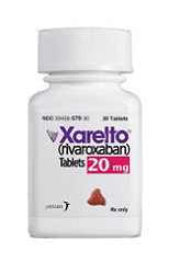
ROME—Preliminary results from the ANNEXA-4 study suggest that andexanet alfa, an investigational antidote to factor Xa inhibitors, can be effective in patients with acute major bleeding.
The drug reversed the anticoagulant effects of rivaroxaban, apixaban, and enoxaparin in this study, providing “excellent” or “good” hemostatic efficacy in 79% of patients over 12 hours.
Thrombotic events occurred in 18% of patients, and 15% died during the 30-day follow-up period.
According to investigators, these events occurred within the range expected in this patient population, given the severity of their bleeding, their underlying thrombotic risk, and the low percentage of patients who restarted anticoagulant therapy following their bleeding episode.
Stuart J. Connolly, MD, of McMaster University in Hamilton, Ontario, Canada, presented results from ANNEXA-4 at ESC Congress 2016 (abstract 5718).
Results were also published in NEJM. The study was funded by Portola Pharmaceuticals Inc.
Patients and treatment
The preliminary analysis of the phase 3/4 ANNEXA-4 trial included 67 patients. All of these patients were evaluated for safety, and 47 were evaluated for efficacy. The mean age of both populations was 77.1, and slightly more than half of the patients were male.
All patients received andexanet alfa given as a bolus dose over 30 minutes, followed by a 2-hour infusion. Patients received a low or high dose depending on which factor Xa inhibitor they received and the time they received it. The patients were evaluated for 30 days following andexanet alfa administration.
The co-primary efficacy endpoints are the percent change in anti-factor Xa activity at 2 hours and the assessment of hemostasis over 12 hours following the infusion. Hemostatic efficacy is assessed by an independent endpoint adjudication committee as excellent, good, or poor/none.
Efficacy
“In this preliminary analysis, [andexanet alfa] was effective in rapidly reversing anti-factor Xa inhibitor activity and restoring normal blood clotting in real-world patients with factor Xa inhibitor-related bleeding,” Dr Connolly said.
Of the 47 patients evaluable for efficacy, 32 were receiving an anticoagulant due to atrial fibrillation, 12 had venous thromboembolism (VTE), and 3 had both atrial fibrillation and VTE.
Twenty-six patients were receiving rivaroxaban, 20 were receiving apixaban, and 1 was receiving enoxaparin. Twenty-five patients had gastrointestinal bleeding, 20 had intracranial bleeding, and 2 had bleeding at other sites.
Forty-two patients received a low dose of andexanet alfa, and 5 received a high dose. The mean time from presentation to the emergency department and the administration of the andexanet alfa bolus was 4.8 ± 1.8 hours.
After the bolus, the median anti-factor Xa activity decreased by 89% from baseline among patients receiving rivaroxaban and by 93% among those receiving apixaban. At the end of the 2-hour infusion, the decrease from baseline was 86% and 92%, respectively.
Twelve hours after the infusion ended, the median anti-factor Xa activity had decreased 64% from baseline among patients receiving rivaroxaban and 31% among those receiving apixaban.
Overall, at 12 hours, clinical hemostasis was rated excellent or good in 79% of patients. Hemostatic efficacy was rated as excellent or good in 81% of the patients on rivaroxaban, 75% of the patients on apixaban, and in the 1 patient on edoxaban.
Safety
Of the 67 patients in the safety population, 47 were receiving an anticoagulant due to atrial fibrillation, 15 had VTE, and 5 had both atrial fibrillation and VTE.
Thirty-two patients were receiving rivaroxaban, 31 were receiving apixaban, and 4 were receiving edoxaban. Thirty-eight patients had gastrointestinal bleeding, 28 had intracranial bleeding, and 6 had bleeding at other sites.
There were no infusion reactions, no antibodies to factors Xa or X, and no neutralizing antibodies to andexanet alfa.
Twelve patients (18%) experienced thrombotic events—1 with myocardial infarction, 5 with stroke, 7 with deep-vein thrombosis, and 1 with pulmonary embolism. (Some patients had more than 1 event.)
“This rate of events is not unexpected, considering the thrombotic potential of the patients and the fact that, in most of them, anticoagulation was discontinued at the time of bleeding and not restarted,” Dr Connolly said.
Four patients had a thrombotic event within 3 days of andexanet alfa treatment, and the rest occurred between 4 days and 30 days.
Eighteen patients (27%) resumed anticoagulant therapy within 30 days. One of the 12 patients with a thrombotic event restarted anticoagulation at a therapeutic dose before the event. One other patient received prophylactic doses of enoxaparin before developing a deep-vein thrombosis.
There were 10 deaths (15%), 6 due to cardiovascular events.
Andexanet alfa development
Andexanet alfa is being developed as a reversal agent for apixaban, rivaroxaban, edoxaban, and enoxaparin. Andexanet alfa is intended to be used when reversal of anticoagulation is needed due to life-threatening or uncontrolled bleeding.
The drug is under review by the US Food and Drug Administration (FDA) and the European Medicines Agency. The FDA recently issued a complete response letter regarding the biologics license application for andexanet alfa.
Portola said it plans to meet with the FDA as soon as possible to resolve the outstanding questions in the letter and determine the appropriate next steps. ![]()
Antiplatelet drugs comparable in patients with AMI

Photo courtesy of AstraZeneca
ROME—The antiplatelet drugs prasugrel and ticagrelor produce similar early results in patients with acute myocardial infarction (AMI) treated with percutaneous coronary intervention (PCI), according to PRAGUE-18, the first randomized, head-to-head comparison of the drugs.
“Our findings confirm previous indirect—non-randomized—comparisons of these 2 drugs, based on analyses of various registries,” said study investigator Petr Widimsky MD, DSc, of Charles University in Prague, Czech Republic.
“Thus, both drugs are very effective and safe and significantly contribute to the excellent outcomes of patients with acute myocardial infarction in modern cardiology.”
Dr Widimsky presented results from PRAGUE-18 at ESC Congress 2016 (abstract 5028). This was an investigator-initiated study, so there was no industry support.
The trial included 1230 AMI patients who were randomized to receive prasugrel (n=634) or ticagrelor (n=596) prior to PCI. There were no significant differences in baseline characteristics between the treatment arms.
Randomization took place immediately after a patient’s arrival to the PCI center. Patients received prasugrel at 60 mg, followed by 10 mg per day (5 mg per day if they were older than 75 or weighed less than 60 kg) for 1 year. Patients received ticagrelor at 180 mg, followed by 90 mg twice a day for 1 year.
The study’s primary endpoint was the occurrence of death, re-infarction, urgent target vessel revascularization, stroke, prolonged hospitalization, or serious bleeding requiring transfusion at 7 days (or discharge if earlier).
The trial was halted prematurely, after an interim analysis showed no significant difference in the rate of the primary endpoint between the prasugrel and ticagrelor arms—4.0% and 4.1%, respectively (odds ratio=0.98, P=0.939).
Likewise, there was no significant difference between the treatment arms for any of the components of the primary endpoint.
The key secondary endpoint was a composite of cardiovascular death, non-fatal myocardial infarction, and stroke within 30 days. There was no significant difference in the rate of this endpoint between the prasugrel and ticagrelor arms—2.7% and 2.5%, respectively (odds ratio=1.06, P=0.864).
“This study did not show any difference between ticagrelor and prasugrel in the early phase of acute myocardial infarction treated by primary PCI,” Dr Widimsky concluded.
He and his colleagues are planning the final follow-up of this study at 1 year, which will be completed in 2017. ![]()
FDA approves new indication for ofatumumab in CLL
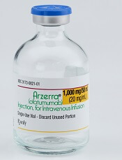
Photo courtesy of GSK
The US Food and Drug Administration (FDA) has approved the use of ofatumumab (Arzerra®) in combination with fludarabine and cyclophosphamide to treat patients with relapsed chronic lymphocytic leukemia (CLL).
Ofatumumab was previously approved by the FDA for use in combination with chlorambucil to treat previously untreated CLL patients who cannot receive fludarabine-based therapy, as monotherapy for CLL that is refractory to fludarabine and alemtuzumab, and as maintenance therapy for patients who are in complete or partial response after receiving at least 2 lines of therapy for recurrent or progressive CLL.
Ofatumumab is a monoclonal antibody designed to target CD20.
The drug’s prescribing information includes a boxed warning noting that hepatitis B virus reactivation can occur in patients receiving CD20-directed cytolytic antibodies, including ofatumumab. In some cases, this results in fulminant hepatitis, hepatic failure, and death.
The boxed warning also states that progressive multifocal leukoencephalopathy, resulting in death, can occur in patients receiving CD20-directed cytolytic antibodies, including ofatumumab.
Ofatumumab is marketed under a collaboration agreement between Genmab and Novartis.
COMPLEMENT 2 trial
The FDA’s latest approval for ofatumumab is based on results of the phase 3 COMPLEMENT 2 trial. Novartis reported top-line results from this study in April.
The trial enrolled 365 patients with relapsed CLL. The patients were randomized 1:1 to receive up to 6 cycles of ofatumumab in combination with fludarabine and cyclophosphamide or up to 6 cycles of fludarabine and cyclophosphamide alone.
The primary endpoint was progression-free survival, as assessed by an independent review committee.
The median progression-free survival was 28.9 months for patients receiving ofatumumab plus fludarabine and cyclophosphamide, compared to 18.8 months for patients receiving fludarabine and cyclophosphamide alone (hazard ratio=0.67, P=0.0032).
Novartis said the safety profile observed in this study was consistent with other trials of ofatumumab, and no new safety signals were observed. ![]()
Study may explain why blood type affects cholera severity
Photo by Daniel Gay
Results of preclinical research may explain why people with blood type O often get more severely ill from cholera than people with other blood types.
The study suggests that, in people with blood type O, cholera toxin hyperactivates a key signaling molecule in intestinal cells.
And high levels of that molecule, cyclic adenosine monophosphate (cAMP), lead to excretion of electrolytes and water—in other words, diarrhea.
“We have shown that blood type influences how strongly cholera toxin activates intestinal cells, leading to diarrhea,” said study author James Fleckenstein, MD, of Washington University School of Medicine in Saint Louis, Missouri.
Dr Fleckenstein and his colleagues reported these findings in The American Journal of Tropical Medicine and Hygiene.
Cholera is caused by Vibrio cholerae, a bacterium that infects cells of the small intestine.
Epidemiologists first noticed 4 decades ago that people with blood type O were more likely to be hospitalized for cholera than people with other blood types, but the reasons for the difference had never been determined.
Although the blood group antigens—A, B, AB, and O—are best known for their presence on red blood cells, they also are found on the surface of many other cell types, including the cells that line the intestine.
To find out what effect cholera toxin has on intestinal cells carrying different blood group antigens, Dr Fleckenstein and his colleagues used clusters of intestinal epithelial stem cells, called enteroids, that can be grown in the lab and differentiated into mature intestinal cells.
The researchers treated 4 groups of enteroids with cholera toxin—2 derived from people with blood type A and 2 from people with blood type O—and measured the amount of cAMP inside the cells. Enteroids from the other 2 blood types—B and AB—were not available at the time the study was done.
The researchers found that levels of cAMP were roughly twice as high in the cells with the type O antigen than in the cells with type A antigen, suggesting that people with type O antigen who were exposed to cholera toxin would suffer more severe diarrhea.
“It is well-established that high levels of this molecule lead to diarrhea, so we’re making the assumption that higher levels lead to even more diarrhea,” said study author F. Matthew Kuhlmann, MD, of Washington University School of Medicine.
“Unfortunately, we have no way directly to link the responses to the volume of diarrhea and, therefore, the severity of disease.”
The researchers confirmed their enteroid results in an intestinal cell line originally derived from a person with blood type A. The cell line was modified to produce the type O antigen instead.
The team found that cholera toxin induced roughly double the amount of cAMP in cells with type O antigen than in those with type A.
Dr Fleckenstein said the researchers are not sure why cholera toxin induces different responses in cells with different blood group antigens on their surfaces.
“The cholera toxin is known to bind weakly to the ABO antigens, so they may be acting as decoys to draw the toxin away from its true target,” Dr Fleckenstein said. “It may be that the type O antigen just isn’t as good of a decoy as the type A antigen.” ![]()
Rule identifies women at low risk of VTE recurrence

of women in a family
ROME—According to researchers, a clinical decision rule can identify women who, after their first unprovoked venous thromboembolism (VTE), have a low risk of VTE recurrence and might safely discontinue anticoagulant therapy.
The researchers evaluated the HERDOO2 rule, which is named after the risk factors the rule employs to determine the likelihood of VTE recurrence, in the REVERSE II trial.
Results from the trial were presented at ESC Congress 2016 (abstract 5721).
According to the HERDOO2 rule, the following risk factors must be considered to determine a patient’s risk of VTE recurrence:
- Hyperpigmentation, Edema, or Redness in either leg
- D-dimer >250 μg/mL on anticoagulants
- Obesity with body mass index >30 kg/m2
- Older than age 65.
Women (but not men) are considered at low risk of VTE recurrence if they have 0 to 1 of these risk factors.
In the REVERSE II trial, researchers tested the HERDOO2 rule in 2779 male and female patients with a first unprovoked VTE who had completed 5 to 12 months of anticoagulant therapy.
After drop-outs and exclusions, 622 women were considered low-risk, based on HERDOO2 criteria, and the majority of these women (n=591) discontinued anticoagulant therapy.
Thirty-one low-risk women continued anticoagulant therapy, as did 1802 men and high-risk women (with 2 or more HERDOO2 criteria). Three hundred and twenty-three men and high-risk women discontinued anticoagulant therapy.
After a year of follow-up, low-risk women who had discontinued anticoagulants had a 3% rate of recurrent VTE per patient year, and low-risk women who continued anticoagulant therapy had no VTEs.
Among the men and high-risk women, the rate of recurrent VTE per patient year was 8.1% in patients who discontinued therapy and 1.6% in patients who continued to receive anticoagulant therapy.
“This is an important finding as, using our rule, over half of women with unprovoked VTE can safely discontinue anticoagulants and be spared the burdens, costs, and risks of life-long anticoagulation,” said study investigator Marc Rodger, MD, of Ottawa Hospital and University of Ottawa in Ontario, Canada.
“Since current consensus guidelines suggest anticoagulants should be continued indefinitely in all patients with unprovoked VTE and non-high bleeding risk, our results are potentially practice-changing.”
Dr Rodger noted, however, that questions remain regarding anticoagulation duration after a first unprovoked VTE.
“One is whether indefinite anticoagulation is required for men and high-risk women, [which] was not the primary focus of our study,” he said. “The second is in the subgroup of post-menopausal women aged 50 and above.”
“In this group, even those who were considered low-risk according to the HERDOO2 rule had a higher than expected rate of recurrent VTE (5.7%) when they discontinued anticoagulants. As such, further validation of HERDOO2 is required in this subset of post-menopausal women.” ![]()

of women in a family
ROME—According to researchers, a clinical decision rule can identify women who, after their first unprovoked venous thromboembolism (VTE), have a low risk of VTE recurrence and might safely discontinue anticoagulant therapy.
The researchers evaluated the HERDOO2 rule, which is named after the risk factors the rule employs to determine the likelihood of VTE recurrence, in the REVERSE II trial.
Results from the trial were presented at ESC Congress 2016 (abstract 5721).
According to the HERDOO2 rule, the following risk factors must be considered to determine a patient’s risk of VTE recurrence:
- Hyperpigmentation, Edema, or Redness in either leg
- D-dimer >250 μg/mL on anticoagulants
- Obesity with body mass index >30 kg/m2
- Older than age 65.
Women (but not men) are considered at low risk of VTE recurrence if they have 0 to 1 of these risk factors.
In the REVERSE II trial, researchers tested the HERDOO2 rule in 2779 male and female patients with a first unprovoked VTE who had completed 5 to 12 months of anticoagulant therapy.
After drop-outs and exclusions, 622 women were considered low-risk, based on HERDOO2 criteria, and the majority of these women (n=591) discontinued anticoagulant therapy.
Thirty-one low-risk women continued anticoagulant therapy, as did 1802 men and high-risk women (with 2 or more HERDOO2 criteria). Three hundred and twenty-three men and high-risk women discontinued anticoagulant therapy.
After a year of follow-up, low-risk women who had discontinued anticoagulants had a 3% rate of recurrent VTE per patient year, and low-risk women who continued anticoagulant therapy had no VTEs.
Among the men and high-risk women, the rate of recurrent VTE per patient year was 8.1% in patients who discontinued therapy and 1.6% in patients who continued to receive anticoagulant therapy.
“This is an important finding as, using our rule, over half of women with unprovoked VTE can safely discontinue anticoagulants and be spared the burdens, costs, and risks of life-long anticoagulation,” said study investigator Marc Rodger, MD, of Ottawa Hospital and University of Ottawa in Ontario, Canada.
“Since current consensus guidelines suggest anticoagulants should be continued indefinitely in all patients with unprovoked VTE and non-high bleeding risk, our results are potentially practice-changing.”
Dr Rodger noted, however, that questions remain regarding anticoagulation duration after a first unprovoked VTE.
“One is whether indefinite anticoagulation is required for men and high-risk women, [which] was not the primary focus of our study,” he said. “The second is in the subgroup of post-menopausal women aged 50 and above.”
“In this group, even those who were considered low-risk according to the HERDOO2 rule had a higher than expected rate of recurrent VTE (5.7%) when they discontinued anticoagulants. As such, further validation of HERDOO2 is required in this subset of post-menopausal women.”
Antiplatelet monitoring doesn’t benefit high-risk patient group

Photo courtesy of NIH
ROME—Results of the ANTARCTIC trial suggest that monitoring platelet function to individualize antiplatelet therapy does not improve outcomes for elderly patients stented for an acute coronary syndrome.
These patients had a high risk of ischemic and bleeding complications, but the study showed no significant difference in the incidence of such complications between patients who were monitored and those who were not.
The findings challenge current international guidelines, which recommend platelet function testing in high-risk patients.
“Platelet function testing is still being used in many centers to measure the effect of antiplatelet drugs and adjust the choice of these drugs and their doses,” said study investigator Gilles Montalescot, MD, PhD, of Hôpital Pitié-Salpêtrière in Paris, France.
“Our study does not support this practice and these recommendations. Although measuring the effect of antiplatelet agents makes sense in order to choose the best
drugs or doses, this costly and more complex strategy does not appear to benefit patients, even when they present with extremely high risk of ischemic and bleeding events like those enrolled in ANTARCTIC.”
Results of the ANTARCTIC trial were presented at ESC Congress 2016 (abstract 2221) and published in The Lancet.
The study was funded by Eli Lilly and Company, Daiichi Sankyo, Stentys, Accriva Diagnostics, Medtronic, and Fondation Coeur et Recherche.
ANTARCTIC enrolled 877 patients, ages 75 and older, who presented with an acute coronary syndrome and underwent coronary stenting.
All patients were started on the antiplatelet agent prasugrel (5 mg), with 442 randomized to the conventional therapy (no adjustment) and 435 randomized to monitoring and treatment adjustment if needed.
Patients in the monitoring arm received 14 days of the daily 5 mg prasugrel dose, then underwent a platelet function test at day 14, followed by medication adjustment if the test showed high or low platelet reactivity. Additional monitoring was performed at day 28 in patients who needed treatment adjustment.
The primary endpoint of the trial was the composite of cardiovascular death, myocardial infarction, stroke, stent thrombosis, urgent revascularization, and bleeding complications at 1 year.
This endpoint occurred at a similar rate in both arms of the study—27.6% in the monitoring arm and 27.8% in the conventional therapy arm (hazard ratio=1.003; P=0.98).
Similarly, there was no significant difference between the arms with regard to the main secondary endpoint—a composite of cardiovascular death, myocardial infarction, stent thrombosis, and urgent revascularization.
This endpoint occurred in 9.9% of patients in the monitoring arm and 9.3% of patients in the conventional arm (hazard ratio=1.06; P=0.80).
“Platelet function monitoring led to a change of treatment in 44.8% of patients who were identified as being over- or under-treated, yet this strategy did not improve ischemic or safety outcomes,” Dr Montalescot noted.
“ANTARCTIC confirms the ARCTIC study in a different population with a different drug and has addressed the potential limitations of the ARCTIC study but finally reached the same conclusion. I expect there will be adjustments of guidelines and practice in light of this.”
BSIs costly for pediatric transplant, cancer patients

Staphylococcus infection
Photo by Bill Branson
Ambulatory bloodstream infections (BSIs) can be costly in young cancer patients and recipients of hematopoietic stem cell transplants, according to research published in Pediatric Blood & Cancer.
Among the 61 patients studied, the median cost for an ambulatory BSI was $40,852, and the median length of hospital stay was 7 days.
For patients who were hospitalized for BSI and other medical issues, the cost and length of stay were much higher.
“This issue has resonance beyond the pediatric stem cell transplant and oncology patient population,” said study author Amy Billett, MD, of the Dana–Farber Cancer Institute and Boston Children’s Hospital in Massachusetts.
“At a time when many aspects of care are being shifted to the home and of heightened attention to safety and cost, this is the new frontier. What we learn about preventing outpatient bloodstream infections in these patients could have broad relevance.”
To determine the economic and hospitalization impact of ambulatory BSIs, Dr Billet and her colleagues retrospectively analyzed data on outpatient BSIs at Dana-Farber/Boston Children’s that occurred between January 1, 2012, and December 31, 2013, and resulted in hospitalization.
The team identified 74 BSIs in 61 patients. Sixty-nine percent of these infections were classified as central-line-associated bloodstream infections.
In 43% of BSIs, the patient’s central line had to be surgically removed. In 15% of cases, the child was transferred to the intensive care unit. Four patients died during hospitalization, and 3 of these deaths were associated with the infections.
Most of the hospitalizations analyzed—62—were due solely to BSIs. The remainder involved at least 1 other medical issue.
The median total cost of BSIs was $40,852, and the median length of hospital stay was 7 days.
The median cost was $36,611 among patients who were hospitalized for BSIs alone (n=62) and $89,935 for patients who were hospitalized for other medical issues as well. The median lengths of hospital stay were 6 days and 15 days, respectively.
The top 3 drivers of cost for all BSIs were room and board (43%), non-chemotherapy medications (22%), and procedures (11%).
Room and board accounted for 42% of charges among patients who were hospitalized for BSIs alone and 44% among the other patients. Non-chemotherapy medications accounted for 20% and 25%, respectively. And procedures accounted for 11% and 10%, respectively.
“Behind these metrics are real and serious risks to patients’ health,” said study author Chris Wong, MD, of Dana-Farber/Boston Children’s.
“The bottom line is that the dollar cost and lengthy hospital stays signal complications that could become life-threatening or delay treatment of the children’s cancer. Reducing these infections is important both for cost containment and quality of care.”
Team uncovers potential treatments for Zika virus

Photo courtesy of
Muhammad Mahdi Karim
Researchers say they have identified compounds that might be used to inhibit Zika virus replication and reduce the ability of the virus to kill brain cells.
The compounds include emricasan (a drug being investigated as a treatment to reduce liver damage from hepatitis C virus), niclosamide (a drug approved in the US to combat parasitic infections), and an investigational cyclin-dependent kinase inhibitor known as PHA-690509.
The researchers described the anti-Zika activity of these compounds in Nature Medicine.
About the virus
The Zika virus has been reported in 60 countries and territories worldwide. Currently, there are no vaccines or effective treatments for the virus.
Research and anecdotal evidence have suggested infection with the Zika virus is related to fetal microcephaly, an abnormally small head resulting from an underdeveloped and/or damaged brain. The virus has also been linked with neurological diseases such as Guillain-Barré syndrome in infected adults.
The Zika virus is spread primarily through bites from infected Aedes aegypti mosquitoes, but it can also be transmitted from mother to child, through sexual contact, via blood transfusion, and possibly through other methods.
“The Zika virus poses a global health threat,” said study author Anton Simeonov, PhD, of the National Center for Advancing Translational Sciences in Bethesda, Maryland.
“While we await the development of effective vaccines, which can take a significant amount of time, our identification of repurposed small-molecule compounds may accelerate the translational process of finding a potential therapy.”
“It takes years, if not decades, to develop a new drug,” noted study author Hongjun Song, PhD, of Johns Hopkins University School of Medicine in Baltimore, Maryland. “In this sort of global health emergency, we don’t have that kind of time.”
“So instead of using new drugs, we chose to screen existing drugs,” added Guo-li Ming, MD, PhD, also of Johns Hopkins. “In this way, we hope to create a therapy much more quickly.”
Identifying potential treatments
The researchers screened 6000 compounds, both investigational and approved (in the US), looking for drugs that might be effective against the Zika virus.
The team first exposed cell cultures to 3 strains of the virus—Ugandan, Cambodian, and Puerto Rican. Then, they introduced the various compounds and looked for indicators of cell death.
The researchers identified more than 100 promising compounds. The 3 lead compounds were emricasan, niclosamide, and PHA-690509.
These compounds were effective either in inhibiting the replication of Zika or in preventing the virus from killing brain cells. Emricasan prevents cell death, while niclosamide and PHA-690509 stop virus replication.
The researchers found that combining emricasan and PHA-690509 prevented both cell death and virus replication.
Dr Song cautioned that the 3 drugs “are very effective against Zika in the dish, but we don’t know if they can work in humans in the same way.”
For example, he noted that, although niclosamide can safely treat parasites in the human gastrointestinal tract, researchers have not yet determined if the drug can penetrate the central nervous system of adults or a fetus inside a carrier’s womb to treat the brain cells targeted by Zika.
Furthermore, it’s not clear if the drugs would address the wide range of effects of Zika infection.
“To address these questions, additional studies need to be done in animal models as well as humans to demonstrate their ability to treat Zika infection,” Dr Ming said. “So we could still be years away from finding a treatment that works.”