User login
Method identifies effective treatments for leukemias, lymphomas
An ex vivo drug screening method can reveal optimal therapies for patients with hematologic malignancies, according to research published in The Lancet Haematology.
Researchers used a method called pharmacoscopy to measure single-cell responses to possible treatments in samples from patients with leukemias and lymphomas.
The team then used these results to guide treatment decisions and found that pharmacoscopy-guided treatment greatly improved response rates and progression-free survival (PFS).
“Having a robust, fast, and reliable predictive test at our disposal during the patient treatment process, especially at the time of relapse where a new intervention must be selected quickly, will change how medical doctors prioritize drugs to use for late-stage patients,” said study author Philipp Staber, MD, of Medical University of Vienna in Austria.
With pharmacoscopy, hundreds of drug options can be pre-tested ex vivo in small liquid biopsy samples collected from individual patients. The effects of each drug on the individual cells are quantified using high-throughput and high-content automated confocal microscopy.
In combination with specially developed analysis methods, machine learning, and other algorithms, pharmacoscopy allows quantification of never-before visualized phenotypes. The method was first described last April in Nature Chemical Biology.
Now, Dr Staber and his colleagues have reported, in The Lancet Haematology, an interim analysis of the first clinical trial testing pharmacoscopy-guided treatment.
There were 17 evaluable patients, all of whom had aggressive hematologic malignancies. This included diffuse large B-cell lymphoma (n=6), acute myeloid leukemia (n=3), B-cell acute lymphoblastic leukemia (n=2), precursor B-cell lymphoblastic lymphoma (n=1), peripheral T-cell lymphoma (n=1), primary mediastinal B-cell lymphoma (n=1), T-cell lymphoblastic lymphoma (n=1), follicular lymphoma (n=1), and T-cell prolymphocytic leukemia (n=1).
The researchers compared outcomes with pharmacoscopy-guided treatment to outcomes with the most recent regimen on which the patient had progressed.
The overall response rate was 88% with pharmacoscopy-guided treatment and 24% with the patients’ most recent previous treatment regimen (odds ratio=24.38; 95%, CI 3.99–125.4; P=0.0013).
None of the patients had progressive disease as their best overall response when they received pharmacoscopy-guided treatment. However, 7 patients had progressive disease in response to their most recent prior regimen.
At the time of analysis, 8 patients (47%) still had ongoing responses after pharmacoscopy-guided treatment.
In addition, pharmacoscopy-guided treatment significantly improved PFS. The median PFS was 22.6 weeks with pharmacoscopy-guided treatment and 5.7 weeks with the most recent prior regimen (hazard ratio=3.14; 95%, CI 1.37–7.22; P=0.0075).
“Evidence that the pharmacoscopy approach is helpful for clinical evaluation of therapy is wonderful,” said study author Giulio Superti-Furga, PhD, of CeMM Research Center for Molecular Medicine in Vienna, Austria.
“Single-cell functional analysis of primary material gives unprecedented resolution and precision that we are sure to further develop in the future to address yet more diseases.” ![]()
PTSD can persist in cancer survivors
Cancer patients may experience lasting post-traumatic stress disorder (PTSD), according to a study published in the journal Cancer.
Approximately one-fifth of patients involved in the study experienced PTSD several months after their cancer diagnosis, and roughly a third of these patients continued to live with PTSD 4 years later.
Researchers say these findings highlight the need for early identification, careful monitoring, and treatment of PTSD in cancer survivors.
Caryn Mei Hsien Chan, PhD, of the National University of Malaysia in Kuala Lumpur, and her colleagues conducted this research.
The study included 469 adults with various cancers who were within 1 month of cancer diagnosis at enrollment.
Patients who had significant psychological distress (defined as a Hospital Anxiety and Depression Scale total cutoff score of 16 or higher) underwent
testing for PTSD at 6 months of follow-up. All patients were tested for PTSD at 4 years of follow-up (regardless of their Hospital Anxiety and Depression Scale score).
The incidence of PTSD was 21.7% at 6 months and 6.1% at 4 years. Although overall rates of PTSD decreased with time, roughly one-third of patients initially diagnosed with PTSD were found to have persistent or worsening symptoms 4 years later.
“Many cancer patients believe they need to adopt a ‘warrior mentality’ and remain positive and optimistic from diagnosis through treatment to stand a better chance of beating their cancer,” Dr Chan said.
“To these patients, seeking help for the emotional issues they face is akin to admitting weakness. There needs to be greater awareness that there is nothing wrong with getting help to manage the emotional upheaval—particularly depression, anxiety, and PTSD—post-cancer.”
Dr Chan also stressed that many patients live in fear that their cancer may come back, and they may think the cancer has returned with every lump or bump, pain or ache, fatigue or fever.
In addition, cancer survivors might skip visits to their oncologists or other physicians to avoid triggering memories of their past cancer experience. This can lead to delays in seeking help for new symptoms or even refusal of treatment for unrelated conditions.
“We need psychological evaluation and support services for patients with cancer at an initial stage and at continued follows-up because psychological well-being and mental health—and by extension, quality of life—are just as important as physical health,” Dr Chan noted. ![]()
Withdrawn drug receives orphan designation for HA
The US Food and Drug Administration (FDA) has granted orphan drug designation to rofecoxib (TRM-201) as a potential treatment for degenerative joint disease in hemophilia, also known as hemophilic arthropathy (HA).
Rofecoxib is a COX-2 selective non-steroidal anti-inflammatory drug (NSAID) that was previously sold in the US under the name Vioxx.
Vioxx was FDA-approved to relieve the signs and symptoms of osteoarthritis, manage acute pain in adults, and treat primary dysmenorrhea.
Merck & Co. pulled Vioxx from the US market in 2004 due to safety concerns. The drug was shown to increase a person’s risk of cardiovascular events, including heart attack and stroke.
Now, Tremeau Pharmaceuticals, Inc., is working to bring rofecoxib back to market to treat patients with HA.
HA patients should not receive traditional NSAIDs due to their effects on platelet aggregation and the risk of gastrointestinal ulcers associated with these drugs. Therefore, high potency opioids are the current standard of care in HA.
“Being granted an orphan drug designation for rofecoxib by FDA is an important regulatory milestone for Tremeau and affirms our strategy of providing non-opioid pain treatments for rare diseases like hemophilic arthropathy,” said Bradford C. Sippy, chief executive officer of Tremeau.
Sippy is a former Merck employee who helped with the recall of Vioxx and knew the final patent protecting the drug’s monopoly was expiring this fall.
When it stopped making Vioxx, Merck was facing thousands of lawsuits from people claiming the drug caused their heart attacks or strokes.
Merck’s own research showed the drug doubled those risks, but lawyers for patients claimed the company downplayed or concealed that. Merck initially fought the lawsuits but, in 2007, agreed to a $4.85 billion settlement.
If approved to treat HA, rofecoxib would carry a warning about the increased risk of heart attack and stroke associated with the drug.
Although the orphan designation for rofecoxib is a step toward FDA approval, Sippy said Tremeau must still raise $25 million or more to fund trials of the drug in hemophilia patients.
About orphan designation
The FDA grants orphan designation to products intended to treat, diagnose, or prevent diseases/disorders that affect fewer than 200,000 people in the US.
The designation provides incentives for sponsors to develop products for rare diseases. This may include tax credits toward the cost of clinical trials, prescription drug user fee waivers, and 7 years of market exclusivity if the product is approved. ![]()
Nilotinib approved to treat kids with CML in EU
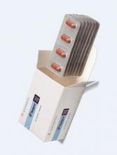
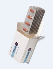
The European Commission has approved nilotinib (Tasigna®) for the treatment of pediatric patients.
The drug is now approved to treat children age 2 and older with newly diagnosed, Philadelphia chromosome-positive (Ph+), chronic phase (CP) chronic myeloid leukemia (CML) or Ph+ CP-CML with resistance or intolerance to prior therapy, including imatinib.
Nilotinib is the only second-generation tyrosine kinase inhibitor currently approved in the European Union (EU) for the treatment of Ph+ CP-CML in children. The approval applies to all EU member states.
According to Novartis, the expanded indication for nilotinib is based on 2 prospective studies of the drug in children with Ph+ CP-CML, which were part of a formal “pediatric investigation plan” agreed upon with the European Medicines Agency.
The company said 69 patients received nilotinib in these studies. The patients ranged in age from 2 to 18. They had either newly diagnosed Ph+ CP-CML or Ph+ CP-CML with resistance or intolerance to prior therapy, including imatinib.
In the newly diagnosed patients, the major molecular response (MMR) rate was 60.0% (95% CI: 38.7, 78.9) at 12 cycles, with 15 patients achieving MMR.
In patients with resistance or intolerance to prior therapy, the MMR rate was 40.9% (95% CI: 26.3, 56.8) at 12 cycles, with 18 patients being in MMR.
In newly diagnosed patients, the cumulative MMR rate was 64.0% by cycle 12. In patients with resistance or intolerance to prior therapy, the cumulative MMR rate was 47.7% by cycle 12.
Adverse events were generally consistent with those observed in adults, with the exception of hyperbilirubinemia and transaminase elevation, which were reported at a higher frequency than in adults.
The rate of grade 3/4 hyperbilirubinemia was 13.0%, the rate of grade 3/4 AST elevation was 1.4%, and the rate of grade 3/4 ALT elevation was 8.7%.
There were no deaths on treatment or after treatment discontinuation. ![]()
CD22-CAR therapy shows activity in rel/ref B-ALL
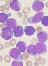
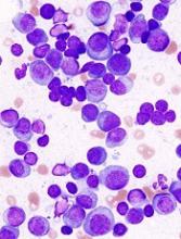
Researchers say they have reported the first results demonstrating clinical activity of a CD22-directed chimeric antigen receptor (CAR) T-cell therapy in B-cell acute lymphoblastic leukemia (B-ALL).
The team conducted a phase 1 study of the therapy in 21 children and adults with relapsed/refractory B-ALL.
Twelve patients achieved a complete response (CR) to the treatment, with 3 patients still in CR at last follow-up.
Sixteen patients developed cytokine release syndrome (CRS), all grade 1 or 2.
Crystal Mackall, MD, of Stanford University in California, and her colleagues reported these results in Nature Medicine.*
“This is the first time that we’ve seen response rates anything like we achieved when we were first testing the CD19 CAR T therapy,” Dr Mackall said.
“We were all a little worried that we wouldn’t find anything comparable, but this study gives hope to the idea that there may be another similar, very potent treatment.”
Patients
Dr Mackall and her colleagues studied the CD22-CAR T-cell therapy in 21 patients with relapsed/refractory B-ALL. They had a median age of 19 (range, 7 to 30).
All of the patients had received a hematopoietic stem cell transplant at least once, and 2 patients had 2 prior transplants each. Seventeen patients had received prior CD19-directed immunotherapy. Fifteen had received CD19-directed CAR T-cell therapy, and 2 had received blinatumomab.
Lymphoblasts were CD19− or CD19dim in 10 patients (9 who had received a CD19-CAR and 1 treated with blinatumomab).
The median CD22 site density was 2839 molecules per cell (range, 613 to 13,452).
Dosing and DLTs
Patients received the CD22-CAR T-cell therapy at 1 of 3 dose levels:
- 0.3 × 10⁶ CD22-CAR T cells per kg body weight (n=6)
- 1 × 10⁶ cells per kg (n=13)
- 3 × 10⁶ cells per kg (n=2).
There was 1 dose-limiting toxicity (DLT) at the first dose level. It was grade 3, self-limited, noninfectious diarrhea that occurred during CRS and resolved with supportive care.
The other DLT occurred in a patient who received treatment at the third dose level. This patient had grade 4 hypoxia that was associated with rapid disease progression. The patient required brief intubation, and the hypoxia was resolved within 24 hours of starting steroid treatment.
Based on these results, the second dose level became the recommended phase 2 dose.
Other adverse events
The researchers said the primary toxicity was CRS, which occurred in 16 patients. Nine patients had grade 1 CRS, and 7 had grade 2.
There were no cases of irreversible neurotoxicity or seizure reported. Among the first 16 patients with complete assessments, there were cases of transient visual hallucinations (n=2), mild unresponsiveness (n=1), mild disorientation (n=1), and mild to moderate pain (n=2). However, these incidents resolved by day 28.
One patient died from gram-negative rod sepsis that developed after the resolution of CRS and neutrophil count recovery to >1000 cells/μL blood. The patient had a history of multi-organ failure due to sepsis.
Response
Twelve patients (57%) had a CR, and 9 of them were minimal residual disease negative.
One CR occurred at the lowest dose of therapy, 1 occurred at the highest dose, and the remaining 10 CRs occurred in patients who received dose level 2.
The researchers said there was no evidence to suggest that previous CD19-directed immunotherapy or diminished surface expression of CD19 impacted response to the CD22-CAR T-cell therapy.
Of the 9 patients who did not respond, 4 progressed and 5 had stable disease.
The researchers said 4 non-responders had “very high disease burden with rapid disease progression.” And 2 non-responders expressed diminished or partial CD22 on leukemic blasts at the time of enrollment.
The median duration of response was 6 months (range, 1.5 to 21+ months). Three patients are still in CR at 6, 9, and 21 months of follow-up.
“The take-home message is that we’ve found another CAR T-cell therapy that displays high-level activity in this phase 1 trial,” Dr Mackall said. “But the relapse rate was also high. So this forces the field to get even more sophisticated. How much of a target is needed for successful, long-lasting treatment? What happens if we target both CD19 and CD22 simultaneously?”
The researchers are already tackling the last question by testing a CAR T-cell therapy that recognizes both CD19 and CD22. They’ve confirmed this therapy can kill cancer cells in vitro and in vivo. Now, they’re testing it in a clinical trial that has opened at Stanford University and will open soon at the National Cancer Institute. ![]()
*This research was supported, in part, by the Intramural Research Program, National Cancer Institute and NIH Clinical Center, National Institutes of Health; by a Stand Up to Cancer–St. Baldrick’s Pediatric Dream Team translational research grant; and by a St. Baldrick’s Foundation Scholar Award.
NOACs may do less harm to kidneys than warfarin
New research suggests warfarin may produce worse renal outcomes than non-vitamin K antagonist oral anticoagulants (NOACs) in patients with atrial fibrillation.
In a study of close to 10,000 patients, dabigatran and rivaroxaban were associated with lower risks of adverse renal outcomes than warfarin.
However, risks with warfarin and apixaban were not significantly different.
This research was published in the Journal of the American College of Cardiology.*
“Kidney function decline in patients taking oral anticoagulant drugs is an important topic that has been overlooked in previous clinical trials,” said study author Xiaoxi Yao, PhD, of Mayo Clinic in Rochester, Minnesota.
“Even our past work at Mayo Clinic has been primarily focused on risks for stroke or bleeding.”
For the current study, Dr Yao and her colleagues examined the de-identified records of 9769 patients from the OptumLabs Data Warehouse.
The patients had atrial fibrillation and started taking oral anticoagulants—apixaban, dabigatran, rivaroxaban, or warfarin—between Oct. 1, 2010, and April 30, 2016.
The researchers looked at 4 indicators of kidney function in these patients:
- A 30% or greater decline in estimated glomerular filtration rate (eGFR)
- Doubled serum creatinine level
- Acute kidney injury (AKI)
- Kidney failure.
For the entire study population, the cumulative risk of each event occurring within 2 years of beginning anticoagulation was as follows:
- 24.4% for ≥30% eGFR
- 4.0% for doubled creatinine level
- 14.8% for AKI
- 1.7% for kidney failure.
“Our study demonstrated that renal function decline is very common among atrial fibrillation patients on blood thinners,” Dr Yao said. “About 1 in 4 patients had significantly reduced kidney function within 2 years of being on any of these medications, and 1 in 7 patients had acute kidney injury.”
“In general, patients with atrial fibrillation taking blood-thinning medications tend to have declining kidney function over time,” added study author Peter Noseworthy, MD, of Mayo Clinic in Rochester, Minnesota.
“However, our findings indicate that the non-vitamin K antagonist oral anticoagulants, as a group, are associated with less injury to kidneys than warfarin.”
When the researchers compared all 3 NOACs to warfarin, they found NOAC use was associated with a reduced risk of:
- ≥30% decline in eGFR—hazard ratio (HR)=0.77 (P<0.001)
- Doubling of creatinine—HR=0.62 (P=0.03)
- AKI—HR=0.68 (P<0.001).
However, results differed in one-to-one comparisons.
There was no significant difference between warfarin and apixaban for any of the renal endpoints measured. The HRs (for apixaban vs warfarin) were:
- 0.88 for ≥30% decline in eGFR (P=0.25)
- 0.80 for doubling of creatinine (P=0.51)
- 0.84 for AKI (P=0.16)
- 1.02 for kidney failure (P=0.95).
Dabigatran, on the other hand, was associated with lower risks of ≥30% decline in eGFR and AKI than warfarin. The HRs were as follows:
- 0.72 for ≥30% decline in eGFR (P=0.01)
- 0.64 for doubling of creatinine (P=0.24)
- 0.55 for AKI (P<0.001)
- 0.45 for kidney failure (P=0.21).
Rivaroxaban was associated with lower risks of ≥30% decline in eGFR, doubling of creatinine, and AKI. The HRs were as follows:
- 0.73 for ≥30% decline in eGFR (P<0.001)
- 0.46 for doubling of creatinine (P<0.01)
- 0.69 for AKI (P<0.001)
- 0.63 for kidney failure (P=0.13).
“Patients with atrial fibrillation already face a high risk of kidney disease, perhaps because many such patients have risk factors, such as advanced age, diabetes, and hypertension,” Dr Yao said. “Many drugs these patients are taking rely on kidney function for drug elimination. Therefore, it is particularly important for these patients to choose a drug that minimizes the impact on kidneys.”
“Since non-vitamin K antagonist oral anticoagulants have a different drug mechanism than warfarin, researchers have hypothesized that non-vitamin K antagonist oral anticoagulants may be related to better renal outcomes. Our study is among the first few studies confirming this hypothesis.” ![]()
*This study was funded by the Mayo Clinic Robert D. and Patricia E. Kern Center for the Science of Health Care Delivery, which receives no industry funding. However, study authors did declare industry relationships.
Technique may predict treatment outcomes in MM
A cell-measuring technique can help predict how multiple myeloma (MM) patients will respond to different therapies, according to research published in Nature Communications.
The technique involves a microfluidic device known as a serial suspended microchannel resonator (sSMR).
The sSMR measures cell mass and allows researchers to calculate growth rates of single cells over short periods of time.
The device consists of a series of sensors that weigh cells as they flow through tiny channels.
The sSMR can measure 50 to 100 cells per hour.
Over a 20-minute period, each cell is weighed 10 times. This is enough to get an accurate mass accumulation rate (MAR), which is a measurement of the rate at which the cells gain mass.
With previous work, researchers showed that MAR can reveal drug susceptibility. A decrease in MAR following drug treatment means cells are sensitive to the drug. If cells are resistant, there is no change in MAR.
For the current study, Scott Manalis, PhD, of the Massachusetts Institute of Technology in Cambridge, and his colleagues tested drugs on cells from MM patients.
The team compared MAR results obtained via sSMR to outcomes of treatment in 9 patients.
For each patient, the researchers tracked the cells’ response to 3 different drugs—bortezomib, lenalidomide, and dexamethasone—plus combinations of those drugs.
In all 9 cases, the sSMR results matched the outcomes seen in patients, as measured by clinical biomarkers in the bloodstream.
“When the clinical biomarkers showed that the patients should be sensitive to a drug, we also saw sensitivity by our measurement,” said study author Mark Stevens, PhD, of the Massachusetts Institute of Technology and Dana-Farber Cancer Institute.
“[I]n cases where the patients were resistant, we saw that in the clinical biomarkers as well as our measurement.”
The researchers believe their device could potentially be used in MM patients at the time of relapse when the disease may have developed resistance to specific therapies.
“At this time of relapse, we would take a bone marrow biopsy from a patient, and we would test each therapy individually or in combinations that are typically used in the clinic,” Dr Stevens said. “At that point, we’d be able to inform the clinician as to which therapy or combinations of therapies this patient seems to be most sensitive or most resistant to.”
Now, the researchers are planning to conduct a larger clinical study to validate this approach. They also aim to investigate the possibility of using this technology for other cancers.
Some of the researchers involved in this study are cofounders of Travera and Affinity Biosensors, 2 companies that develop techniques relevant to this research. ![]()
A cell-measuring technique can help predict how multiple myeloma (MM) patients will respond to different therapies, according to research published in Nature Communications.
The technique involves a microfluidic device known as a serial suspended microchannel resonator (sSMR).
The sSMR measures cell mass and allows researchers to calculate growth rates of single cells over short periods of time.
The device consists of a series of sensors that weigh cells as they flow through tiny channels.
The sSMR can measure 50 to 100 cells per hour.
Over a 20-minute period, each cell is weighed 10 times. This is enough to get an accurate mass accumulation rate (MAR), which is a measurement of the rate at which the cells gain mass.
With previous work, researchers showed that MAR can reveal drug susceptibility. A decrease in MAR following drug treatment means cells are sensitive to the drug. If cells are resistant, there is no change in MAR.
For the current study, Scott Manalis, PhD, of the Massachusetts Institute of Technology in Cambridge, and his colleagues tested drugs on cells from MM patients.
The team compared MAR results obtained via sSMR to outcomes of treatment in 9 patients.
For each patient, the researchers tracked the cells’ response to 3 different drugs—bortezomib, lenalidomide, and dexamethasone—plus combinations of those drugs.
In all 9 cases, the sSMR results matched the outcomes seen in patients, as measured by clinical biomarkers in the bloodstream.
“When the clinical biomarkers showed that the patients should be sensitive to a drug, we also saw sensitivity by our measurement,” said study author Mark Stevens, PhD, of the Massachusetts Institute of Technology and Dana-Farber Cancer Institute.
“[I]n cases where the patients were resistant, we saw that in the clinical biomarkers as well as our measurement.”
The researchers believe their device could potentially be used in MM patients at the time of relapse when the disease may have developed resistance to specific therapies.
“At this time of relapse, we would take a bone marrow biopsy from a patient, and we would test each therapy individually or in combinations that are typically used in the clinic,” Dr Stevens said. “At that point, we’d be able to inform the clinician as to which therapy or combinations of therapies this patient seems to be most sensitive or most resistant to.”
Now, the researchers are planning to conduct a larger clinical study to validate this approach. They also aim to investigate the possibility of using this technology for other cancers.
Some of the researchers involved in this study are cofounders of Travera and Affinity Biosensors, 2 companies that develop techniques relevant to this research.
Antimalarial might fight Zika virus
Preclinical research suggests a drug used to prevent and treat malaria may also be effective against Zika virus.
Investigators found that chloroquine can protect human neural progenitors from infection with Zika virus.
The drug also decreased Zika-induced mortality in one mouse model and hindered transmission of Zika virus from mother to fetus in another mouse model.
Alexey Terskikh, PhD, of Sanford Burnham Prebys Medical Discovery Institute in La Jolla, California, and his colleagues conducted this research and disclosed the results in Scientific Reports.
“There is still an urgent need to bolster our preparedness and capacity to respond to the next Zika outbreak,” Dr Terskikh said. “Our latest research suggests the antimalaria drug chloroquine may be an effective drug to treat and prevent Zika infections.”
The investigators first found that chloroquine reduced Zika infection in primary human fetal neural precursor cells.
The team also discovered that chloroquine reduced the percentage of Zika-positive cells and the level of apoptosis in neurospheres derived from human induced pluripotent stem cells.
The investigators then tested chloroquine in AG129 mice, which lack receptors for type I and II interferons and have been used to model Zika virus infection.
Some of these mice received chloroquine (50 mg/kg/day) in their drinking water for 2 days and were then infected with Zika virus. The mice continued to receive chloroquine at the same dose for 5 days and then received 5 mg/kg/day until the end of the experiment.
Control AG129 mice were infected with Zika virus and received regular drinking water.
Compared to controls, chloroquine-treated mice had significantly prolonged survival and a significant reduction in Zika-induced weight loss (P<0.01 for both).
Next, the investigators used SJL mice to study horizontal and vertical transmission of Zika. They observed successful transmission of the virus from males to females and from mothers to pups.
The team then analyzed the effects of chloroquine on fetal transmission of Zika. Pregnant SJL mice were infected with Zika and given drinking water containing chloroquine (30 mg/kg/day) starting on day E13.5. The mice were euthanized on E18.5, and maternal blood and fetal brain samples were collected.
Chloroquine treatment resulted in a roughly 20-fold reduction in Zika virus titers in the maternal blood and fetal brain.
“Although chloroquine didn’t completely clear Zika from infected mice, it did reduce the viral load, suggesting it could limit the neurological damage found in newborns infected by the virus,” Dr Terskikh said.
“Chloroquine has a long history of successfully treating malaria, and there are no reports of it causing birth defects. Additional studies are certainly needed to determine the precise details of how it works. But given its low cost, availability, and safety history, further study in a clinical trial to test its effectiveness against Zika virus in humans is merited.”
Team discovers mechanism of resistance in AML
Researchers say they have uncovered a target to overcome drug resistance in acute myeloid leukemia (AML).
The team discovered how a linkage between 2 proteins enables AML cells to resist chemotherapy and showed that disrupting the linkage could render the cells vulnerable to treatment.
The researchers believe their discovery could lead to drugs to enhance chemotherapy in patients with AML and other cancers.
John Schuetz, PhD, of St. Jude Children’s Research Hospital in Memphis, Tennessee, and his colleagues described this research in Nature Communications.
The team launched their experiments based on previous findings that a protein called ABCC4 was greatly elevated in aggressive cases of AML.
Dr Schuetz and his colleagues searched for other proteins that might interact with ABCC4 and enable its function. The team’s screening of candidate proteins yielded one, MPP1, which was also greatly increased in AML.
The researchers found the 2 proteins are connected, and the connection enables cells to assume the characteristics of highly proliferative leukemia cells.
These experiments involved genetically altering hematopoietic progenitor cells to have high MPP1 and ABCC4 levels. The cells were grown in culture and then replated to see if they would continue to grow, as such self-renewal is a hallmark of leukemia cells.
The researchers found that serial regrowth depended on the cells having high levels of both ABCC4 and MPP1.
“Typically, if you take normal progenitors and you replate, you could do that one time, maybe twice,” Dr Schuetz said. “But our big surprise was that overexpressing MPP1—analogous to what you would see in leukemia—allows those progenitors to self-renew, to be replated over and over, to form new colonies.”
The experiments also revealed that MPP1 and ABCC4 functioned at the cell membrane, where they could play a role in the machinery that would rid the leukemia cells of chemotherapy drugs.
“When we disrupted their interaction, ABCC4 moved off the membrane and the cells became more sensitive to drugs used in AML—drugs that are pumped out of the cell by ABCC4,” Dr Schuetz said.
By screening thousands of compounds, the researchers identified some that could disrupt the ABCC4-MPP1 connection. One, called Antimycin-A, reversed drug resistance in AML cell lines and in cells from AML patients.
Antimycin-A is too toxic to be used in chemotherapy, but the researchers believe identification of the compound should aid the search for other, less-toxic drugs to disrupt the ABCC4-MPP1 interaction.
The team’s findings could also enable clinicians to identify AML patients with high levels of ABCC4 and MPP1. In such patients, drugs that disrupt ABCC4-MPP1 might enhance the effectiveness of standard chemotherapy, Dr Schuetz said.
He also noted that other cancers, including breast and colon cancer and medulloblastoma, show high levels of both ABCC4 and MPP1. Chemotherapy for those cancers might also be enhanced by drugs that disrupt ABCC4-MPP1.
Product approved to treat patients with hemophilia A and inhibitors
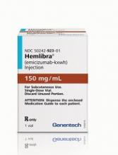
The US Food and Drug Administration (FDA) has approved use of emicizumab-kxwh (Hemlibra®), a bispecific factor IXa- and factor X-directed antibody.
Emicizumab is approved as routine prophylaxis to prevent or reduce the frequency of bleeding episodes in adults and children who have hemophilia A and factor VIII (FVIII) inhibitors.
Emicizumab can be self-administered once-weekly via subcutaneous injection.
The labeling for emicizumab contains a boxed warning noting that patients who received emicizumab in conjunction with activated prothrombin complex concentrate developed thrombotic microangiopathy (TMA) and thromboembolic events (TEs).
Therefore, patients should discontinue prophylactic use of bypassing agents (BPAs) the day before starting prophylaxis with emicizumab.
The FDA granted the approval of emicizumab to Genentech, Inc. The agency granted the application for emicizumab priority review, and the product received breakthrough therapy and orphan drug designations.
Access to emicizumab
According to Genentech, emicizumab will be available shortly.
The company said it will be offering comprehensive services to help minimize barriers to access and reimbursement. Patients can call 866-436-5427 (866-HEMLIBRA) for more information.
For people who qualify, Genentech plans to offer patient assistance programs through Genentech Access Solutions. More information is available at 866-422-2377 (866-4ACCESS) or http://www.Genentech-Access.com.
Emicizumab trials
The biologics license application for emicizumab was supported by results from a pair of phase 3 studies—HAVEN 1 and HAVEN 2.
Results from HAVEN 1 were published in NEJM and presented at the 26th ISTH Congress in July. Interim results from HAVEN 2 were presented at ISTH as well.
HAVEN 1
The study enrolled 109 patients (age 12 and older) with hemophilia A and FVIII inhibitors who were previously treated with BPAs on-demand or as prophylaxis.
The patients were randomized to receive emicizumab prophylaxis or no prophylaxis. On-demand treatment of breakthrough bleeds with BPAs was allowed.
There was a significant reduction in treated bleeds of 87% with emicizumab prophylaxis compared to no prophylaxis (95% CI: 72.3; 94.3, P<0.0001). And there was an 80% reduction in all bleeds with emicizumab (95% CI: 62.5; 89.8, P<0.0001).
Adverse events occurring in at least 5% of patients treated with emicizumab were local injection site reactions, headache, fatigue, upper respiratory tract infection, and arthralgia.
Two patients experienced TEs, and 3 had TMA while receiving emicizumab prophylaxis and more than 100 u/kg/day of activated prothrombin complex concentrate, on average, for 24 hours or more before the event. Two of these patients had also received recombinant factor VIIa.
Neither TE required anticoagulation therapy, and 1 patient restarted emicizumab. The cases of TMA observed were transient, and 1 patient restarted emicizumab.
HAVEN 2
In this single-arm trial, researchers evaluated emicizumab prophylaxis in children younger than 12 years of age who had hemophilia A with FVIII inhibitors.
The interim efficacy analysis, after at least 12 weeks of treatment, included 23 children.
After a median observation time of 38.1 weeks, 87% (95% CI: 66.4; 97.2) of children who received emicizumab experienced 0 treated bleeds. The percentage with 0 treated or non-treated bleeds was lower, at 34.8% (95% CI: 16.4; 57.3).
The most common adverse events (observed in at least 10% of patients) were mild injection site reactions and nasopharyngitis. No TEs or TMAs were observed.
HAVEN 3 and 4
Emicizumab is now being studied in 2 additional phase 3 trials.
In HAVEN 3, researchers are evaluating emicizumab prophylaxis dosed once weekly or once every other week in patients age 12 and older with hemophilia A without FVIII inhibitors.
In HAVEN 4, researchers are evaluating emicizumab prophylaxis dosed every 4 weeks in patients age 12 and older with hemophilia A, with or without inhibitors.


