User login
CHMP endorses drug delivery system
The European Medicines Agency’s Committee for Medicinal Products for Human Use (CHMP) has recommended a label variation for Neulasta® (pegfilgrastim) to include the Neulasta® Onpro® Kit.
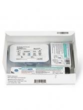
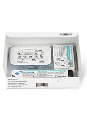
The Neulasta Onpro Kit is an on-body injector (OBI) delivery system for Neulasta, which is approved in the European Union to reduce the duration of neutropenia and the incidence of febrile neutropenia in adults treated with cytotoxic chemotherapy for malignancy (with the exception of chronic myeloid leukemia and myelodysplastic syndromes).
The Neulasta Onpro Kit includes a specifically designed Neulasta pre-filled syringe along with a single-use OBI. The small, lightweight OBI is applied to a patient’s skin on the same day of chemotherapy.
The OBI is intended to facilitate timed delivery of the correct dose of Neulasta and eliminate the need for patients to return to a healthcare setting the day after they receive chemotherapy.
The CHMP’s opinion on the Neulasta Onpro Kit will be reviewed by the European Commission (EC).
If the EC agrees with the CHMP, the centralized marketing authorization of Neulasta will be updated to include information on the Neulasta Onpro Kit in the drug’s label.
This change will be valid throughout the European Union. Norway, Iceland, and Liechtenstein will make corresponding decisions about Neulasta’s label on the basis of the EC’s decision.
The EC typically makes a decision within 67 days of the CHMP’s recommendation.
Team identifies biomarkers for cGVHD in kids
SALT LAKE CITY—Researchers say they have identified prognostic biomarkers for chronic graft-vs-host disease (cGVHD) in children.
The group found that recent thymic emigrants (RTEs) and regulatory T cells expressing CD31 and CD45RA (Treg RTEs) were significantly lower at day 100 after transplant in patients who developed cGVHD.
Because prior research indicated that RTEs are higher in adults with cGVHD, the researchers concluded that thymic function may play a bigger role in cGVHD development for children than for adults.
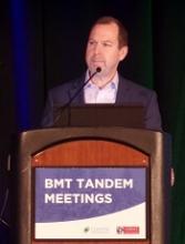
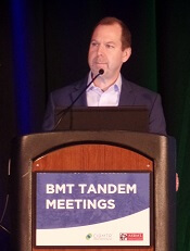
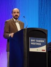
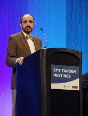
Geoff D.E. Cuvelier, MD, of CancerCare Manitoba in Winnipeg, Canada, presented these findings at the 2018 BMT Tandem Meetings (abstract 104).
“Our group became interested in these populations of recent thymic emigrants when 2 adult studies1,2 . . . showed that higher proportions of recent thymic emigrants at day 100 were prognostic for the development of chronic graft-vs-host disease,” Dr Cuvelier said.
This led his group to evaluate whether RTEs (CD4+CD45RA+CD31+ T cells), as well as Treg RTEs—Tregs (CD4+CD25+CD127Low) that co-express naïve and recently emigrated markers (CD45RA+CD31+)—are prognostic for pediatric cGVHD at day 100 after allogeneic hematopoietic stem cell transplant (allo-HSCT).
The researchers’ study enrolled patients younger than 18 years of age who underwent allo-HSCT to treat malignant and non-malignant diseases. There were 144 patients who had 1 year of follow-up after allo-HSCT.
Thirty-seven of the patients (25.7%) had cGVHD, and 34 (23.6%) had late acute GVHD (post-day 100). The remaining 73 patients (50.7%) had neither cGVHD nor late acute GVHD, so they served as controls.
Twenty cases of cGVHD were severe, 12 were moderate, and 5 were mild. cGVHD was initially diagnosed at a mean of 192 days post-transplant. The median number of organ systems involved was 3 (range, 1-6). Seventeen patients (46%) had overlap syndrome.
The researchers found that RTEs as a percentage of CD4 T cells were significantly lower at day 100 in patients with cGVHD than in controls—7.8% and 13.5%, respectively (P=0.007). The difference between controls and patients with late acute GVHD was not significant—13.5% and 9.4%, respectively (P=0.08).
Treg RTEs as a percentage of all Tregs were also significantly lower at day 100 in patients with cGVHD than in controls—6.4% and 13.2%, respectively (P=0.002). Again, the difference between controls and patients with late acute GVHD was not significant—13.2% and 9.1%, respectively (P=0.06).
However, further analysis revealed that percentages of RTEs and Treg RTEs were significantly lower at day 100 in patients who developed any type of GVHD after day 100.
The percentage of RTEs was 13.5% in controls and 8.5% in patients with any GVHD after day 100 (P=0.009). The percentage of Treg RTEs was 13.2% and 7.6%, respectively (P=0.004).
“Both recent thymic emigrants and Treg RTEs at day 100 are prognostic biomarkers for chronic graft-vs-host disease in children in this large, multi-institutional, prospective trial,” Dr Cuvelier said in closing.
“Unlike adults, in children, RTEs are proportionally lower, not higher, in patients that later develop chronic GVHD compared to no chronic GVHD. This may suggest that thymic dysfunction and thymic output may play a greater role in chronic GVHD development in children compared to adults.”
To expand upon this research, Dr Cuvelier and his colleagues are planning to begin model building using prognostic cellular and plasma biomarkers at day 100 to determine high-risk profiles for cGVHD.
1. Greinix HT et al; CD19+CD21low B Cells and CD4+CD45RA+CD31+ T Cells Correlate with First Diagnosis of Chronic Graft-versus-Host Disease. BBMT 2015; 21(2):250-258.
2. Li AM et al. An Early Naïve T Cell Population Lacking PD1 Expression at Day 100 As A Prognostic Biomarker of Chronic GVHD. TSS 2017; 210.6.
CHMP backs bosutinib for newly diagnosed CML
The European Medicines Agency’s Committee for Medicinal Products for Human Use (CHMP) has recommended expanding the approved use of bosutinib (BOSULIF) to include treatment of patients with newly diagnosed, chronic phase, Philadelphia chromosome-positive (Ph+) chronic myelogenous leukemia (CML).
Bosutinib is currently approved in Europe to treat patients with Ph+ CML in chronic, accelerated, or blast phase who have received one or more tyrosine kinase inhibitors and for whom imatinib, nilotinib, and dasatinib are not considered appropriate treatment options.
The CHMP’s opinion on bosutinib will be reviewed by the European Commission (EC). If the EC agrees with the CHMP, the commission will grant a centralized marketing authorization that will be valid in the European Union. Norway, Iceland, and Liechtenstein will make corresponding decisions on the basis of the EC’s decision.
The EC typically makes a decision within 67 days of the CHMP’s recommendation.
The CHMP’s recommendation for bosutinib is based on results from the BFORE trial, which were recently published in the Journal of Clinical Oncology.
The publication included data on 536 patients newly diagnosed with chronic phase CML. They were randomized 1:1 to receive bosutinib (n=268) or imatinib (n=268).
The modified intent-to-treat population included Ph+ patients with e13a2/e14a2 transcripts who had at least 12 months of follow-up. In this group, there were 246 patients in the bosutinib arm and 241 in the imatinib arm.
In the modified intent-to-treat population, the rate of major molecular response at 12 months was 47.2% in the bosutinib arm and 36.9% in the imatinib arm (P=0.02). The rate of complete cytogenetic response was 77.2% and 66.4%, respectively (P<0.008).
In the entire study population, 22.0% of patients receiving bosutinib and 26.8% of those receiving imatinib discontinued treatment—12.7% and 8.7%, respectively, due to drug-related toxicity.
Adverse events that were more common in the bosutinib arm than the imatinib arm included grade 3 or higher diarrhea (7.8% vs 0.8%), increased alanine levels (19% vs 1.5%), increased aspartate levels (9.7% vs 1.9%), cardiovascular events (3% vs 0.4%), and peripheral vascular events (1.5% vs 1.1%).
Cerebrovascular events were more common with imatinib than bosutinib (0.4% and 0%, respectively).
CHMP supports approval of denosumab in MM
The European Medicines Agency’s Committee for Medicinal Products for Human Use (CHMP) has recommended expanding the current indication for denosumab (XGEVA®).
The CHMP said the drug should be approved for use in preventing skeletal related events (SREs) in adults with advanced malignancies involving bone, which includes multiple myeloma (MM).
Denosumab is currently approved in Europe to prevent SREs—defined as radiation to bone, pathologic fracture, surgery to bone, and spinal cord compression—in adults with bone metastases from solid tumors.
The drug is also approved for the treatment of adults and skeletally mature adolescents with giant cell tumor of bone that is unresectable or where surgical resection is likely to result in severe morbidity.
The CHMP’s opinion on expanding the indication for denosumab will be reviewed by the European Commission (EC).
If the EC agrees with the CHMP, the commission will grant a centralized marketing authorization that will be valid in the European Union. Norway, Iceland, and Liechtenstein will make corresponding decisions on the basis of the EC’s decision.
The EC typically makes a decision within 67 days of the CHMP’s recommendation.
The CHMP’s recommendation for denosumab is based on data from the ’482 study, which were recently published in The Lancet Oncology.
In this phase 3 trial, denosumab proved non-inferior to zoledronic acid for delaying SREs in patients with newly diagnosed MM and bone disease.
Researchers randomized 1718 patients to receive subcutaneous denosumab at 120 mg and intravenous placebo every 4 weeks (n=859) or intravenous zoledronic acid at 4 mg (adjusted for renal function at baseline) and subcutaneous placebo every 4 weeks (n=859). All patients also received investigators’ choice of first-line MM therapy.
The median time to first on-study SRE was 22.8 months for patients in the denosumab arm and 24 months for those in the zoledronic acid arm (hazard ratio=0.98; 95% confidence interval: 0.85-1.14; P non-inferiority=0.010).
There were fewer renal treatment-emergent adverse events in the denosumab arm than the zoledronic acid arm—10% and 17%, respectively.
But there were more hypocalcemia adverse events in the denosumab arm than the zoledronic acid arm—17% and 12%, respectively.
Expanded UCB product can stand alone
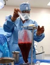
SALT LAKE CITY—The expanded umbilical cord blood (UCB) product NiCord can be used as a stand-alone graft, according to research presented at the 2018 BMT Tandem Meetings.
Researchers found that a single NiCord unit provided “robust” engraftment in a phase 1/2 study of patients with high-risk hematologic malignancies.
NiCord recipients had quicker neutrophil and platelet engraftment than matched control subjects who received standard myeloablative UCB transplant (single or double).
Mitchell Horwitz, MD, of the Duke University Medical Center in Durham, North Carolina, presented these results at the meeting as abstract 49.* The research was sponsored by Gamida Cell, the company developing NiCord.
“[NiCord] is an ex vivo expanded cell product that’s derived from an entire unit of umbilical cord blood,” Dr Horwitz explained. “It’s manufactured starting with a CD133-positive selection, which is the progenitor cell population that’s cultured, and a T-cell containing CD133-negative fraction that is provided also at the time of transplant.”
“The culture system contains nicotinamide—that’s the active ingredient in the culture. And that’s supplemented with cytokines—thrombopoietin, IL-6, FLT-3 ligand, and stem cell factor. The culture is 21 days.”
Previous research showed that double UCB transplant including a NiCord unit could provide benefits over standard double UCB transplant. This led Dr Horwitz and his colleagues to wonder if NiCord could be used as a stand-alone graft.
So the team evaluated the safety and efficacy of NiCord alone in 36 adolescents/adults with high-risk hematologic malignancies.
Patients had acute myelogenous leukemia (n=17), acute lymphoblastic leukemia (n=9), myelodysplastic syndrome (n=7), chronic myelogenous leukemia (n=2), and Hodgkin lymphoma (n=1).
Most patients had intermediate (n=15) or high-risk (n=13) disease. They had a median age of 44 (range, 13-63) and a median weight of 75 kg (range, 41-125).
Treatment
For conditioning, 19 patients received thiotepa, busulfan, and fludarabine. Fifteen patients received total body irradiation and fludarabine with or without cyclophosphamide or thiotepa. And 2 patients received clofarabine, fludarabine, and busulfan.
Most patients had a 4/6 human leukocyte antigen (HLA) match (n=26), 8 had a 5/6 HLA match, and 2 had a 6/6 HLA match.
The median total nucleated cell dose was 2.4 x 10⁷/kg prior to expansion of the UCB unit and 3.7 x 10⁷/kg after expansion. The median CD34+ cell dose was 0.2 x 10⁶/kg and 6.3 x 10⁶/kg, respectively.
“CD34 cells were expanded 33-fold in the 3-week culture system,” Dr Horwitz noted. “That translated to a median CD34 dose of 6.3 x 10⁶/kg, a dose comparable to what would be obtained from an adult donor graft.”
Engraftment
There was 1 case of primary graft failure and 2 cases of secondary graft failure. One case of secondary graft failure was associated with an HHV-6 infection, and the other was due to a lethal adenovirus infection.
Of those patients who engrafted, 97% achieved full donor chimerism, and 3% had mixed chimerism.
Dr Horwitz and his colleagues compared engraftment results in the NiCord recipients to results in a cohort of patients from the CIBMTR registry who underwent UCB transplants from 2010 to 2013. They had similar characteristics as the NiCord patients—age, conditioning regimen, disease status, etc.
In total, there were 148 CIBMTR registry patients, 20% of whom received a single UCB unit.
The median time to neutrophil engraftment was 11.5 days (range, 6-26) with NiCord and 21 days in the CIBMTR matched control cohort (P<0.001). The cumulative incidence of neutrophil engraftment was 94.4% and 89.7%, respectively.
The median time to platelet engraftment was 34 days (range, 25-96) with NiCord and 46 days in the CIBMTR controls (P<0.001). The cumulative incidence of platelet engraftment was 80.6% and 67.1%, respectively.
“There’s a median 10-day reduction in neutrophil recovery [and] 12-day reduction in time to platelet recovery [with NiCord],” Dr Horwitz noted. “There is evidence of robust and durable engraftment with a NiCord unit, with one patient now over 7 years from his first transplant on the pilot trial.”
Relapse, survival, and GVHD
Dr Horwitz reported other outcomes in the NiCord recipients without making comparisons to the CIBMTR matched controls.
The estimated 2-year rate of non-relapse mortality in NiCord recipients was 23.8%, and the estimated 2-year incidence of relapse was 33.2%.
The estimated disease-free survival was 49.1% at 1 year and 43.0% at 2 years. The estimated overall survival was 51.2% at 1 year and 2 years.
At 100 days, the rate of grade 2-4 acute GVHD was 44.0%, and the rate of grade 3-4 acute GVHD was 11.1%.
The estimated 1-year rate of mild to severe chronic GVHD was 40.5%, and the estimated 2-year rate of moderate to severe chronic GVHD was 9.8%.
Dr Horwitz said these “promising results” have led to the launch of a phase 3 registration trial in which researchers are comparing NiCord to standard single or double UCB transplant. The trial is open for accrual.
*Information in the abstract differs from the presentation.
Minihaplo-BMT can cure severe hemoglobinopathies
SALT LAKE CITY—Cure of severe hemoglobinopathies is now possible for most patients, according to a speaker at the 2018 BMT Tandem Meetings.
Results of a small study suggest that nonmyeloablative haploidentical bone marrow transplant (minihaplo-BMT), when used with 400 cGy total body irradiation and post-transplant cyclophosphamide, can cure sickle cell disease (SCD) and beta-thalassemia while conferring limited toxicity.
“This is our second study showing that you can certainly get rid of these conditions using haploidentical donors and, also, that you can cure these conditions with a nonmyeloablative regimen,” said Javier Bolaños-Meade, MD, of Johns Hopkins University in Baltimore, Maryland.
“What we saw [in the current study] is that increasing the total body irradiation from 200 cGy to 400 cGy can overcome the very high rate of graft failure we observed in our prior study.”
However, the results also suggest that major ABO incompatible donors should be avoided.
Dr Bolaños-Meade presented these findings as a late-breaking abstract (LBA3) at this year’s BMT Tandem Meetings.
He began the presentation by pointing out that allogeneic BMT is the only potential cure for SCD and beta-thalassemia. Unfortunately, most of these patients have been excluded from transplant due to a lack of matched donors, concerns over graft-vs-host disease (GVHD), and/or compromised end organ function precluding them from myeloablative conditioning.
With a prior study, Dr Bolaños-Meade and his colleagues found that minihaplo-BMT with post-transplant cyclophosphamide expanded the donor pool and was well tolerated in patients with SCD. However, the rate of secondary graft failure was high, at 43%.
“We hypothesized that increasing the amount of radiation that was given to these patients, making the regimen absolutely more intensive, will help us to overcome this rate of graft failure and retain the tolerability of the regimen,” Dr Bolaños-Meade said.
He and his colleagues tested this theory in 12 patients with SCD and 5 with beta-thalassemia.
The SCD patients had a median age of 26 (range, 6-31), and most (n=8) were female. Ten were African-American, and 2 were of Arab descent.
All SCD patients had at least 2 hospital admissions in the last 2 years, 5 had osteonecrosis, 3 were transfusion-dependent, 2 had a history of acute chest syndrome, and 1 patient had changes in brain magnetic resonance imaging.
The thalassemia patients had a median age of 7 (range, 6-16). Four were female, and all 5 were of Arab descent. All patients were transfusion-dependent from infancy.
Treatment
All patients received antithymocyte globulin (rabbit) at 0.5 mg/kg on day -9 and 2 mg/kg on days -8 and -7, fludarabine at 30 mg/m2 on days -6 to -2, cyclophosphamide at 14.5 mg/kg on days -6 and -5, and total body irradiation at 400 cGy on day -1.
Patients received unmanipulated bone marrow that was collected and infused on day 0. Donors included 1 aunt, 4 brothers, 3 sisters, 5 mothers, and 4 fathers. There were 2 major ABO incompatible pairs, 5 minor ABO incompatible pairs, and 10 ABO compatible pairs.
For GVHD prophylaxis, patients received 50 mg/kg/day of cyclophosphamide on days 3 and 4, sirolimus (levels of 5-10 ng/dl) from days 5 to 365, and mycophenolate mofetil at 1 g every 8 hours from days 5 to 35.
Efficacy
All 17 patients are still alive at a median follow-up of 15 months (range, 3-34).
“Of all the patients transplanted, we only observed 1 graft failure,” Dr Bolaños-Meade said. “This is in sharp contrast to the 40% graft failure in our prior study.”
Thirteen patients achieved full donor chimerism, and 3 had mixed chimerism.
Four of the 5 beta-thalassemia patients achieved full donor chimerism, and 1 had mixed chimerism. All 5 patients became transfusion-independent.
Eleven of the 12 SCD patients engrafted, and 9 achieved full donor chimerism.
All but one of the engrafted SCD patients became transfusion-independent. The exception was a mixed chimeric patient who had a major ABO mismatched donor. The patient remains transfusion-dependent more than 30 months after BMT.
“This is the main problem we have seen in all our sickle cell and hemoglobinopathy experiences,” Dr Bolaños-Meade said. “If you have a major ABO incompatibility, and there’s mixed chimerism, you’re asking for trouble.”
Toxicity
All SCD patients had sickle cell crises after antithymocyte globulin. Other toxicities included sirolimus-induced diabetes in 1 patient, worsening of Meniere’s disease in 1 patient, and BK cystitis in 1 patient.
Two patients developed grade 2 acute GVHD, and 1 developed grade 3 acute GVHD. Three patients had chronic GVHD, 2 mild and 1 moderate.
However, all cases of GVHD resolved. And, of the 8 engrafted patients with more than 1 year of follow-up, 6 are off systemic immunosuppression.
Regimen deemed ‘safe and feasible’ in MM
SALT LAKE CITY—A novel transplant regimen is “safe and feasible” for patients with multiple myeloma (MM), according to a presentation at the 2018 BMT Tandem Meetings.
Researchers conducted a phase 1 trial investigating the addition of elotuzumab and autologous peripheral blood mononuclear cell (PBMC) reconstitution to standard autologous hematopoietic stem cell transplant (ASCT) and lenalidomide maintenance in MM patients.
The regimen was considered well tolerated, as most adverse events (AEs) were grade 1 or 2.
The complete response (CR) rate was 40% at both 90 days and 1 year after ASCT.
Keren Osman, MD, of Mount Sinai Health System in New York, New York, reported these results as abstract 26.* This is an investigator-sponsored study, conducted in collaboration with Bristol Myers Squibb, the company marketing elotuzumab.
Dr Osman noted that elotuzumab is a humanized IgG1 monoclonal antibody targeting SLAMF7. The drug directly activates natural killer (NK) cells through the SLAMF7 pathway and Fc receptors. Elotuzumab also targets SLAMF7 on MM cells and facilitates interaction with NK cells to mediate the killing of MM cells through antibody-dependent cellular cytotoxicity.
“The hypothesis behind this phase 1 study was that . . ., in the peri-transplant period, if we add elotuzumab, we will succeed in promoting NK-mediated elimination of residual multiple myeloma cells as well as promote transition from innate to adaptive immune responses against multiple myeloma-associated antigens,” Dr Osman said.
She and her colleagues tested this hypothesis in 15 MM patients. They had a median age of 59 (range, 49-70), and 60% of them were male.
Forty percent of patients had stage I disease, 27% had stage II, and 20% had stage III. All patients had an ECOG status of 0 (47%) or 1 (53%).
The patients had a median of 174 days since diagnosis (range, 98-393).
All patients had received 1 to 2 prior lines of therapy. They all had bortezomib as part of their induction therapy, and 67% of them had received lenalidomide.
Seventy-three percent of patients had any cytogenetic abnormality, and 47% had high-risk cytogenetics.
Treatment
Patients underwent leukopheresis for PBMC collection, followed by standard peripheral blood stem cell mobilization and harvest.
They received standard melphalan conditioning on day -1 and autologous stem cell rescue on day 0. Autologous PBMCs were reinfused on day 3 after stem cell infusion.
Elotuzumab was given at 20 mg/kg intravenously on day 4. The drug was given every 28 days up to cycle 12.
Lenalidomide maintenance was given at 10 mg orally on days 1 to 21 of every 28-day cycle, starting with cycle 4 of elotuzumab (90 days post-ASCT). Maintenance could continue beyond cycle 12 at the investigators’ discretion.
Thirteen patients completed a year of treatment. One patient withdrew from the study due to early progression. The other patient withdrew due to personal choice and had a very good partial response (VGPR) at withdrawal.
Safety
“The majority of the adverse events, including infusion reactions attributable to elotuzumab, were grade 2 or lower,” Dr Osman noted. “And there were no AEs related to PBMC administration.”
One patient had a delay in hematopoietic reconstitution, which resulted in hospitalization exceeding 21 days.
One patient had grade 3 hypertension, which was attributed to elotuzumab and resolved with supportive care.
Eight patients had grade 3 lymphopenia, which was associated with elotuzumab, during the maintenance phase. Patients received prophylactic antibiotics, and there were no opportunistic infections.
Efficacy
At screening (after induction but before ASCT), 13 patients (87%) had a VGPR, and 2 (13%) had a partial response (PR).
At 90 days post-ASCT, 2 patients (13%) had a stringent CR, and 4 (27%) had a CR. Six patients (40%) had a VGPR, and 2 (13%) had a PR. One patient (7%) had progressed.
“In this very high-risk PR/VGPR population, stem cell transplant with elotuzumab and PBMC infusion resulted in a CR rate of 40%—with 13% of patients achieving stringent CR—by day 90,” Dr Osman noted. “And maintenance elotuzumab plus lenalidomide promoted further conversion to 33% stringent CR by 1 year.”
At 1 year, 5 patients (33%) had a stringent CR, and 1 (7%) had a CR. Six patients (40%) had a VGPR, and 1 (7%) had a PR. Two patients (13%) had progressed.
So the 1-year progression-free survival rate was 86% (13/15).
“In conclusion, we see that elotuzumab and PBMC administration with standard autologous stem cell transplant and lenalidomide maintenance for consolidation therapy in multiple myeloma is certainly both safe and feasible,” Dr Osman said. “We’re planning a phase 2 study.”
*Information in the abstract differs from the presentation.
Exercise doesn’t offset VTE risk from TV viewing
Getting the recommended amount of physical activity does not eliminate the increased risk of venous thromboembolism (VTE) associated with frequent TV viewing, according to research published in the Journal of Thrombosis and Thrombolysis.
In a study of more than 15,000 Americans, people who reported watching TV “very often” had nearly twice the risk of VTE as people who said they “never or seldom” watched TV.
People who watched TV very often had an increased VTE risk even when researchers adjusted for physical activity levels, smoking status, body mass index, and other factors.
“These results suggest that even individuals who regularly engage in physical activity should not ignore the potential harms of prolonged sedentary behaviors such as TV viewing,” said study author Yasuhiko Kubota, of the University of Minnesota in Minneapolis.
“Avoiding frequent TV viewing, increasing physical activity, and controlling body weight might be beneficial to prevent VTE.”
Kubota and his colleagues came to these conclusions after analyzing data from the Atherosclerosis Risk in Communities (ARIC) Study, an ongoing, population-based, prospective study conducted in the US.
The researchers evaluated 15,158 Americans who were 45 to 64 years of age when ARIC started in 1987.
Study subjects were initially asked about their health status, whether they exercised or smoked, and whether they were overweight or not. Over the years, ARIC team members have been in regular contact with subjects to ask about any hospital treatment they might have received.
When subjects were asked about their TV viewing habits, most said they watched TV “sometimes” (n=7094). Fewer subjects said they watched TV “often” (n=4023), “never or seldom” (n=2815), and “very often” (n=1226).
Results
Via hospital records and imaging tests, Kubota and his colleagues identified 691 cases of VTE in the study cohort.
In assessing the risk of VTE, the researchers compared the “never or seldom” TV viewing group to the other viewing groups.
In a model adjusted for age, sex, and race, “sometimes” viewers had a hazard ratio (HR) of VTE of 1.17, the “often” group had an HR of 1.31, and the “very often” group had an HR of 1.88.
In a second model, the researchers adjusted for the aforementioned factors as well as smoking status, leisure-time physical activity, eGFR, and prevalent cardiovascular disease.
In this model, “sometimes” viewers had an HR of VTE of 1.16, the “often” group had an HR of 1.26, and the “very often” group had an HR of 1.71.
In a third model, Kubota and his colleagues adjusted for all of the aforementioned factors as well as body mass index.
In this model, “sometimes” viewers had an HR of VTE of 1.13, the “often” group had an HR of 1.20, and the “very often” group had an HR of 1.53.
The researchers noted that subjects who met the American Heart Association’s recommended level of physical activity but watched TV “very often” had an HR of VTE of 1.80. Subjects with a poor physical activity level who watched TV “very often” had an HR of 2.07.
Kubota and his colleagues also found that obese subjects had an increased risk of VTE, and TV viewing appeared to add to that risk.
Obese subjects who watched TV “very often” had an HR of VTE of 3.70. For obese subjects who “never or seldom” watched TV, the HR was 1.90.
Cognitive impairment in HSCT recipients
New research suggests the risk of cognitive impairment after hematopoietic stem cell transplant (HSCT) is greatest among recipients of myeloablative allogeneic (allo) HSCTs.
Compared to healthy controls, patients who received myeloablative allo-HSCT had a significantly higher risk of global cognitive deficit at a few time points after transplant.
There was a trend toward increased global cognitive deficit in recipients of allo-HSCT who had reduced-intensity conditioning (RIC), but there was no increased risk of global cognitive deficit in recipients of autologous (auto) HSCT.
Researchers reported these findings in the Journal of Clinical Oncology.
“With this research from our longitudinal prospective assessment, we are able to deduce that a significant population of allogeneic [HSCT] survivors will experience cognitive impairment that can and will impact different aspects of their lives moving forward,” said study author Noha Sharafeldin, MD, PhD, of the University of Alabama at Birmingham.
“And it’s critical that we as clinicians develop interventions for these patients. This research is just the beginning of our figuring out how we can best care for [HSCT] survivors and enable them to live healthy lives.”
This study included 477 patients who underwent HSCT between 2004 and 2014. There were 236 auto-HSCTs, 128 RIC allo-HSCTs, and 113 myeloablative allo-HSCTs.
Patients underwent standardized neuropsychological testing before HSCT as well as at 6 months, 1 year, 2 years, and 3 years after transplant.
There were 429 patients who completed pre-HSCT testing (89.9%), 341 (81.6%) who underwent testing at 6 months after HSCT, 308 (81.5%) at 1 year, 247 (80.7%) at 2 years, and 227 (81.4%) at 3 years.
Testing was conducted on 8 cognitive domains—executive function, verbal fluency and speed, processing speed, working memory, visual and auditory memory, and fine motor dexterity.
The researchers conducted this testing in 99 healthy control subjects as well.
Before and after HSCT
Prior to HSCT, there were no significant differences in the cognitive domains tested between auto-HSCT recipients and controls or between RIC allo-HSCT recipients and controls.
Recipients of myeloablative allo-HSCT had significantly lower pre-HSCT scores for processing speed (P=0.001), as compared to controls.
After HSCT, there were no significant differences in overall scores between auto-HSCT recipients and controls or between RIC allo-HSCT recipients and controls.
However, recipients of myeloablative allo-HSCT had significantly lower scores than controls for executive function, verbal speed, processing speed, auditory memory, and fine motor dexterity (P≤0.003 for all).
Outcomes over time
For auto-HSCT recipients, there was a significant improvement from pre-HSCT to 6 months post-HSCT in verbal fluency (P<0.001). Meanwhile, there was a significant decrease in verbal processing and fine motor dexterity (P<0.001 for both).
At 3 years, auto-HSCT recipients had a significant increase in verbal fluency (P<0.001) but a significant decrease in visual memory (P=0.001) and fine motor dexterity (P<0.001).
For RIC allo-HSCT recipients, there was a significant decrease from pre-HSCT to 3 years post-HSCT in executive functioning (P=0.003), verbal fluency (P<0.001), and working memory (P<0.001). There were no significant differences between pre-HSCT and 6-month scores.
For patients who received myeloablative allo-HSCT, the only significant difference from pre-HSCT to 6 months or 3 years was a decrease in fine motor dexterity (P<0.001 for both time points).
Global cognitive deficit
There was no significant difference in the prevalence of global cognitive deficit between auto-HSCT recipients and controls before HSCT (22.5% vs 17.2%; P=0.28) or at any time point after—6 months (26.1% vs 16.5%; P=0.07), 1 year (21.4% vs 19.5%; P=0.73), 2 years (21.1% vs 16.4%; P=0.43), and 3 years (18.7% vs 8.7%, P=0.11).
There was no significant difference in the prevalence of global cognitive deficit between RIC allo-HSCT recipients and controls before HSCT (17.2% for both; P=1.0), at 6 months (22.0% vs 16.5%; P=0.35), 1 year (24.1% vs 19.5%; P=0.46), or 2 years (30.6% vs 16.4%; P=0.05) after HSCT.
However, the prevalence was significantly higher for RIC allo-HSCT recipients 3 years after HSCT (35.4% vs 8.7%; P=0.0012).
There was no significant difference in the prevalence of global cognitive deficit between myeloablative allo-HSCT recipients and controls before HSCT (22.3% vs 17.2%; P=0.37) or at 1 year after (28.4% vs 19.5%; P=0.20).
However, the prevalence was significantly higher for myeloablative allo-HSCT recipients at 6 months (31.1% vs 16.5%; P=0.03), 2 years (34.6% vs 16.4%; P=0.02), and 3 years after HSCT (36.0% vs 8.7%; P=0.0015).
“From this data, it’s clear that we have to make strides in supporting allogeneic [HSCT] recipients in their recovery to ensure that we are educating patients and their families on signs of cognitive impairment,” Dr Sharafeldin said. “This data will help us identify patients at highest risk of cognitive impairment and inform the development of interventions that facilitate a patient’s recovery and return to normal life.”
AYA cancer patients lost to follow-up
ORLANDO—Many adolescent and young adult (AYA) cancer survivors don’t continue with follow-up care, according to a new study.
Researchers analyzed nearly 2400 AYAs diagnosed with cancer between 2000 and 2015 and found that 37% of these patients had no follow-up visits since 2015.
The proportion of patients with follow-up visits in 2016 did not vary according to cancer type or insurance status, but it did vary according to patients’ time since last cancer treatment.
These findings were presented at the 2018 Cancer Survivorship Symposium (abstract 29).
“Many adolescents and young adults are unaware of what their long-term risks are after they have finished their cancer treatment,” said study investigator Lynda M. Beaupin, MD, of the Roswell Park Cancer Institute in Buffalo, New York.
“Doctors and other healthcare providers need to be more diligent in letting these patients know about future potential side effects and health risks that could occur based on certain aspects of their cancer treatment.”
In a previous study, Dr Beaupin and her colleagues conducted a focus group discussion with 27 AYAs, ages 18 to 39, to query them about the major barriers that prevented them from seeking follow-up care.
Among the factors mentioned were poor communication with their oncologists, ongoing problems in adjusting to life as a cancer survivor, and loss of health insurance.
As a follow-up to that focus group session, the investigators looked at a larger group of AYAs who were treated at Roswell Park Comprehensive Cancer Center.
The team analyzed data from the cancer center’s tumor registry, which included information on a patient’s current age, age at cancer diagnosis, gender, date of diagnosis, date of most recent doctor visit, and type of cancer.
The investigators also looked at patient-provided information on follow-up doctor appointments and insurance status for 2 cohorts of AYA cancer survivors. Patients in cohort A were diagnosed with cancer between 2010 and 2014, and patients in cohort B were diagnosed between 2005 and 2009.
The most common types of cancer were leukemia/lymphoma, melanoma, germ cell tumors, and thyroid and breast cancers.
Findings
There were 2367 patients, ages 18 to 39, who were diagnosed with cancer between 2000 and 2015.
Thirty-seven percent of these patients did not have a follow-up visit since 2015.
There was no difference in follow-up contact according to cancer type or insurance status. However, length of time since patients’ final cancer treatment was a factor in not scheduling a follow-up appointment.
Regardless of insurance status, 33% of cohort A (diagnosed 2010-2014) and 48% of cohort B (diagnosed 2005-2009) did not have a follow-up visit in 2016.
Among insured patients, 33% of those in cohort A and 47% of those in cohort B did not have a visit in 2016. Among uninsured patients, the percentages were 39% and 46%, respectively.
Next steps
The investigators hope to conduct future studies looking at other factors that may affect AYAs’ willingness to seek follow-up care, such as employment status, distance to a cancer center, whether they are getting tested or treated at a facility other than a cancer center, and how AYAs perceive their current quality of life.
“These patients have the potential to live a normal lifespan, and we need to educate them to become their own advocates so they may receive follow-up care on a regular basis,” Dr Beaupin said.
“We hope they continue to receive that follow-up at an established cancer center that has the facilities to assess cardiac health and provide rehabilitation if needed. There are now established survivorship programs nationwide that can provide follow-up care for those who have completed treatment.”
This study was supported by the National Cancer Institute.
