User login
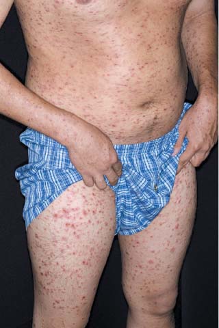
The physician diagnosed pityriasis lichenoides et varioliformis acuta (PLEVA) by clinical appearance and then performed a punch biopsy to confirm the diagnosis.
PLEVA usually presents as a papulonecrotic eruption, but may have vesicles resembling varicella. It can be thought of as “chickenpox that lasts for weeks to months” because new crops of lesions continue to appear. Always suspect PLEVA when a case of “chickenpox” is not going away. A punch biopsy is helpful in making the diagnosis.
PLEVA usually involves the anterior trunk, flexor surfaces of the upper extremities, and the axilla. The eruptions can occur in crops of vesicles that can become hemorrhagic over several weeks or months.
The general health of the patient is unaffected, although most have lymphadenopathy. CD8 T-cells are the predominant cell-type in lesional skin. It is seen more frequently in young men and can go on to become chronic (pityriasis lichenoides chronica).
Various reports suggest the efficacy of macrolides and tetracyclines, probably more for their antiinflammatory properties than for their antibacterial effects. The physician prescribed a one-month course of oral tetracycline 500 mg 4 times daily and the patient’s skin cleared.
Photos and text for Photo Rounds Friday courtesy of Richard P. Usatine, MD. This case was adapted from: Hara J, Usatine R. Other bullous diseases. In: Usatine R, Smith M, Mayeaux EJ, et al, eds. The Color Atlas of Family Medicine. New York, NY: McGraw-Hill; 2009:799-805.
To learn more about The Color Atlas of Family Medicine, see:
* http://www.amazon.com/Color-Atlas-Family-Medicine/dp/0071474641
The Color Atlas of Family Medicine is also available as an app for mobile devices. See