User login
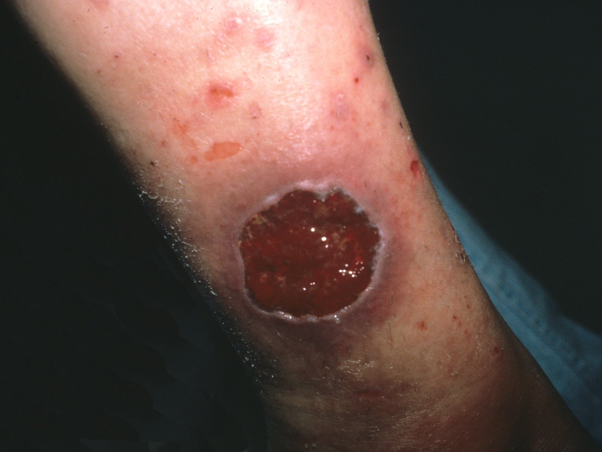
The family physician made the presumptive diagnosis of pyoderma gangrenosum. At least 50% of cases are associated with systemic diseases such as inflammatory bowel disease, hematologic malignancy, and arthritis. It’s estimated that 30% of patients with pyoderma gangrenosum experience pathergy (initiation at the site of trauma or injury). This patient’s history was consistent with pathergy.
Typically pyoderma gangrenosum presents with a deep, painful ulcer. The ulcer has a well-defined border, which is usually violet or blue. The ulcer edge is often undermined and the surrounding skin is erythematous and indurated. Pyoderma gangrenosum usually starts as small papules that coalesce; the central area then undergoes necrosis to form a single ulcer. The lesions are painful and the pain can be severe. Patients may have malaise, arthralgia, and myalgia. Pyoderma gangrenosum is most commonly seen on the legs, but can occur on any skin surface (including the genitalia) and around a stoma.
The physician performed a punch biopsy including the active ulcer and the border to rule out other causes of ulcerative skin lesions. The biopsy showed a neutrophilic infiltrate consistent with pyoderma gangrenosum.
Treatment options include:
- topical medications, such as potent steroid ointments, tacrolimus ointment, and silver sulfadiazine
- intralesional injections with corticosteroids
- systemic treatment with oral corticosteroids
- treatment with mycophenolate mofetil, oral tacrolimus, dapsone, azathioprine, or infliximab (each used successfully, according to published case reports).
Surgical debridement is contraindicated since pathergy will make the lesions worse.
In this case, the patient chose topical clobetasol ointment. She was lost to follow-up.
Photos and text for Photo Rounds Friday courtesy of Richard P. Usatine, MD. This case was adapted from: Flowers D, Usatine R. Pyoderma gangrenosum. In: Usatine R, Smith M, Mayeaux EJ, et al, eds. The Color Atlas of Family Medicine. New York, NY: McGraw-Hill; 2009:735-739.
To learn more about The Color Atlas of Family Medicine, see:
* http://www.amazon.com/Color-Atlas-Family-Medicine/dp/0071474641
* http://www.mhprofessional.com/product.php?isbn=0071474641