User login
A 50-year-old Caucasian man came to the dermatology clinic with a tender growth on his great toe. He first noticed it a year earlier after sustaining trauma to the area. The growth had been getting bigger and had intermittently bled. It had also oozed, so his primary care physician treated it as an infection with topical antibiotics, followed by several courses of oral antibiotics.
Despite therapy, the lesion continued to grow and the patient was referred to us. The patient had a 3.4 cm × 1.8 cm exophytic erythematous nodule on the left lateral great toe (FIGURE).
What is your diagnosis?
How would you manage this condition?
FIGURE
Erythematous nodule on left great toe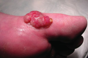
Diagnosis: Acral lentiginous melanoma
A wedge biopsy of the lesion showed that the patient had acral lentiginous melanoma. ALM is a less common subtype of melanoma. (The other subtypes are superficial spreading malignant melanoma, nodular melanoma, and lentigo maligna melanoma.) Acral refers to its typical location, occurring in the distal extremities. The term lentiginous refers to its characteristic histopathologic finding.1,2 Unlike the other subtypes of melanoma, ALM typically affects all ethnic groups. In non-white populations it constitutes 29% to 72% of all melanomas.3 In Caucasian patients it accounts for 2% to 8% of all melanomas.
In many cases, ALM presents with the typical “ABCD” features of melanoma. These include asymmetry, border irregularity, color variegation, and a diameter greater than 6 mm. Lesions on the palmo-plantar surface may be macular, papular, or nodular, and may ulcerate. ALM may involve the nail as well.
As in our case, however, not all cases are typical. Amelanotic lesions may mimic a variety of skin conditions and pose a diagnostic challenge.
ALM is not linked to sun exposure
The pathogenesis of ALM is unclear. Unlike the other types of melanoma, in which sun exposure is clearly identified as an etiologic factor, ALM has not been definitively linked to exposure. In fact, ALM occurs in patients with more darkly pigmented skin, a group not typically considered at risk for melanoma. In African American patients, Hispanic patients, and Asian patients, ALM is the most common subtype of melanoma.4
BRAF/NRAS mutations seem to play an important role in all the major subtypes of cutaneous melanoma.5 Recent studies indicate that nevoid precursor lesions, high total body nevus count, and plantar nevi act as predisposing lesions to ALM.2,6
Injury to the hands and feet. Another common association with ALM is penetrative injury of the hands and feet. There is no clinical or research-based evidence to support this association and therefore the role of trauma in the pathogenesis of ALM is controversial. This association may be seen in some cases because acral sites are more prone to trauma than the rest of the body.7
Trauma leading to hemorrhage mimics ALM
The differential diagnosis includes both melanocytic lesions and non-melanocytic lesions. Benign nevi or dysplastic nevi are included in the first category. Among non-melanocytic lesions, trauma leading to hemorrhage may mimic ALM.2 ALM has also been misdiagnosed as pyogenic granuloma, ischemic ulceration, wart, a foreign body, callus, a nonhealing wound, or other benign skin lesions such as seborrheic keratosis.2,8,9
Amelanotic lesions are particularly misleading on the foot, as they may be frequently misdiagnosed as hyperkeratotic benign lesions.8 Bleeding, itching, satellite lesions, and sudden growth, however, should raise your suspicion of melanoma.10
Excision is best, plus sentinel node mapping
The most effective treatment for ALM is excision—either conservatively or with wide margins—along with sentinel lymph node mapping.11 Systemic chemotherapy is more palliative than curative, as it has not been shown to improve the number of recurrences.2 Patients require lifelong regular full body skin exams with early biopsy of any suspicious skin lesions.9
The prognosis of ALM is poorer than other subtypes of melanoma, mainly because of delayed diagnosis.2,9 The single most important and accurate prognostic factor is the depth of invasion of the tumor as measured by Breslow thickness.10
Other factors that are predictive of a poorer prognosis in ALM patients are male sex and amelanosis.12 It’s important to recognize that the palms, soles, and nails may be sites for melanoma—despite the lack of direct sun exposure.
Our patient required amputation
Our patient’s toe was amputated and the melanoma had a Breslow thickness of 7 mm. Sentinel lymph node biopsy was negative. Our patient received adjuvant chemotherapy with interferon alfa-2B and was followed by oncology.
If you said the face or the arms, you’d be wrong. Given that increased sun exposure and a history of multiple sunburns are recognized as important risk factors for melanoma, it’s easy to assume that the most common sites for melanoma would be exposed areas of the body, such as the face and arms.
Interestingly, though, superficial spreading malignant melanoma—the most common type of malignant melanoma13—is most often found on the legs in women and on the back in men.3,10 One theory is that repeated intense exposure to sunlight may be a more important risk factor than continuous sun exposure.14,15
Correspondence
Rajani Katta, MD, 6620 Main Street, Suite 1425, Houston, TX 77030.
1. Freedberg I. Acral lentiginous melanoma. In: Fitzpatrick’s Dermatology in General Medicine. 5th ed. New York: McGraw-Hill; 1999;767:1081-1083.
2. Stalkup JR, Orengo IF, Katta R. Controversies in acral lentiginous melanoma. Dermatol Surg. 2002;28:1051-1059.
3. Swetter SM. Malignant melanoma. Available at: http://www.emedicine.com/DERM/topic257.htm. Accessed June 6, 2008.
4. Cress RD, Holly EA. Incidence of cutaneous melanoma among non-Hispanic whites, Hispanics, Asians, and blacks: an analysis of California cancer registry data, 1988-93. Cancer Causes Control. 1997;8:246-252.
5. Saldanha G, Potter L, Daforno P, Pringle JH. Cutaneous melanoma subtypes show different BRAF and NRAS mutation frequencies. Clin Cancer Res. 2006;12:4499-4505.
6. Green A, McCredie M, MacKie R, et al. A casecontrol study of melanomas of the soles and palms (Australia and Scotland). Cancer Causes Control. 1999;10:21-25.
7. Kaskel P, Kind P, Sander S, Peter RU, Krähn G. Trauma and melanoma formation: a true association? Br J Dermatol. 2000;143:749-753.
8. Soon SL, Solomon AR, Jr, Papadopoulos D, Murray DR, McAlpine B, Washington CV. Acral lentiginous melanoma mimicking benign disease: the Emory experience. J Am Acad Dermatol. 2003;48:183-188.
9. Rosen T. Acral lentiginous melanoma misdiagnosed as verruca plantaris: a case report. Dermatology Online Journal. 2006;12(4):3.-Available at: http://dermatology.cdlib.org/124/case_reports/melanoma/rosen.html. Accessed June 6, 2008.
10. Bruce AJ, Brodland DG. Overview of skin cancer detection and prevention for the primary care physician. Mayo Clin Proc. 2000;75:491-500.
11. Lang PG. Current concepts in the management of patients with melanoma. Am J Clin Dermatol. 2002;3:401-426.
12. Phan A, Touzet S, Dalle S, Ronger-Savle S, Balme B, Thomas L. Acral lentiginous melanoma: a clinicoprognostic study of 126 cases. Br J Dermatol. 2006;155:561-569.
13. Kang S, Barnhill RL, Mihm MC, Jr, et al. Multiple primary cutaneous melanomas. Cancer. 1992;70:1911-1916.
14. Elwood JM. Melanoma and sun exposure: contrasts between intermittent and chronic exposure. World J Surg. 1992;16:157-165.
15. Elwood JM, Gallagher RP. Body site distribution of cutaneous malignant melanoma in relationship to patterns of sun exposure. Int J Cancer. 1998;78:276-280.
A 50-year-old Caucasian man came to the dermatology clinic with a tender growth on his great toe. He first noticed it a year earlier after sustaining trauma to the area. The growth had been getting bigger and had intermittently bled. It had also oozed, so his primary care physician treated it as an infection with topical antibiotics, followed by several courses of oral antibiotics.
Despite therapy, the lesion continued to grow and the patient was referred to us. The patient had a 3.4 cm × 1.8 cm exophytic erythematous nodule on the left lateral great toe (FIGURE).
What is your diagnosis?
How would you manage this condition?
FIGURE
Erythematous nodule on left great toe
Diagnosis: Acral lentiginous melanoma
A wedge biopsy of the lesion showed that the patient had acral lentiginous melanoma. ALM is a less common subtype of melanoma. (The other subtypes are superficial spreading malignant melanoma, nodular melanoma, and lentigo maligna melanoma.) Acral refers to its typical location, occurring in the distal extremities. The term lentiginous refers to its characteristic histopathologic finding.1,2 Unlike the other subtypes of melanoma, ALM typically affects all ethnic groups. In non-white populations it constitutes 29% to 72% of all melanomas.3 In Caucasian patients it accounts for 2% to 8% of all melanomas.
In many cases, ALM presents with the typical “ABCD” features of melanoma. These include asymmetry, border irregularity, color variegation, and a diameter greater than 6 mm. Lesions on the palmo-plantar surface may be macular, papular, or nodular, and may ulcerate. ALM may involve the nail as well.
As in our case, however, not all cases are typical. Amelanotic lesions may mimic a variety of skin conditions and pose a diagnostic challenge.
ALM is not linked to sun exposure
The pathogenesis of ALM is unclear. Unlike the other types of melanoma, in which sun exposure is clearly identified as an etiologic factor, ALM has not been definitively linked to exposure. In fact, ALM occurs in patients with more darkly pigmented skin, a group not typically considered at risk for melanoma. In African American patients, Hispanic patients, and Asian patients, ALM is the most common subtype of melanoma.4
BRAF/NRAS mutations seem to play an important role in all the major subtypes of cutaneous melanoma.5 Recent studies indicate that nevoid precursor lesions, high total body nevus count, and plantar nevi act as predisposing lesions to ALM.2,6
Injury to the hands and feet. Another common association with ALM is penetrative injury of the hands and feet. There is no clinical or research-based evidence to support this association and therefore the role of trauma in the pathogenesis of ALM is controversial. This association may be seen in some cases because acral sites are more prone to trauma than the rest of the body.7
Trauma leading to hemorrhage mimics ALM
The differential diagnosis includes both melanocytic lesions and non-melanocytic lesions. Benign nevi or dysplastic nevi are included in the first category. Among non-melanocytic lesions, trauma leading to hemorrhage may mimic ALM.2 ALM has also been misdiagnosed as pyogenic granuloma, ischemic ulceration, wart, a foreign body, callus, a nonhealing wound, or other benign skin lesions such as seborrheic keratosis.2,8,9
Amelanotic lesions are particularly misleading on the foot, as they may be frequently misdiagnosed as hyperkeratotic benign lesions.8 Bleeding, itching, satellite lesions, and sudden growth, however, should raise your suspicion of melanoma.10
Excision is best, plus sentinel node mapping
The most effective treatment for ALM is excision—either conservatively or with wide margins—along with sentinel lymph node mapping.11 Systemic chemotherapy is more palliative than curative, as it has not been shown to improve the number of recurrences.2 Patients require lifelong regular full body skin exams with early biopsy of any suspicious skin lesions.9
The prognosis of ALM is poorer than other subtypes of melanoma, mainly because of delayed diagnosis.2,9 The single most important and accurate prognostic factor is the depth of invasion of the tumor as measured by Breslow thickness.10
Other factors that are predictive of a poorer prognosis in ALM patients are male sex and amelanosis.12 It’s important to recognize that the palms, soles, and nails may be sites for melanoma—despite the lack of direct sun exposure.
Our patient required amputation
Our patient’s toe was amputated and the melanoma had a Breslow thickness of 7 mm. Sentinel lymph node biopsy was negative. Our patient received adjuvant chemotherapy with interferon alfa-2B and was followed by oncology.
If you said the face or the arms, you’d be wrong. Given that increased sun exposure and a history of multiple sunburns are recognized as important risk factors for melanoma, it’s easy to assume that the most common sites for melanoma would be exposed areas of the body, such as the face and arms.
Interestingly, though, superficial spreading malignant melanoma—the most common type of malignant melanoma13—is most often found on the legs in women and on the back in men.3,10 One theory is that repeated intense exposure to sunlight may be a more important risk factor than continuous sun exposure.14,15
Correspondence
Rajani Katta, MD, 6620 Main Street, Suite 1425, Houston, TX 77030.
1. Freedberg I. Acral lentiginous melanoma. In: Fitzpatrick’s Dermatology in General Medicine. 5th ed. New York: McGraw-Hill; 1999;767:1081-1083.
2. Stalkup JR, Orengo IF, Katta R. Controversies in acral lentiginous melanoma. Dermatol Surg. 2002;28:1051-1059.
3. Swetter SM. Malignant melanoma. Available at: http://www.emedicine.com/DERM/topic257.htm. Accessed June 6, 2008.
4. Cress RD, Holly EA. Incidence of cutaneous melanoma among non-Hispanic whites, Hispanics, Asians, and blacks: an analysis of California cancer registry data, 1988-93. Cancer Causes Control. 1997;8:246-252.
5. Saldanha G, Potter L, Daforno P, Pringle JH. Cutaneous melanoma subtypes show different BRAF and NRAS mutation frequencies. Clin Cancer Res. 2006;12:4499-4505.
6. Green A, McCredie M, MacKie R, et al. A casecontrol study of melanomas of the soles and palms (Australia and Scotland). Cancer Causes Control. 1999;10:21-25.
7. Kaskel P, Kind P, Sander S, Peter RU, Krähn G. Trauma and melanoma formation: a true association? Br J Dermatol. 2000;143:749-753.
8. Soon SL, Solomon AR, Jr, Papadopoulos D, Murray DR, McAlpine B, Washington CV. Acral lentiginous melanoma mimicking benign disease: the Emory experience. J Am Acad Dermatol. 2003;48:183-188.
9. Rosen T. Acral lentiginous melanoma misdiagnosed as verruca plantaris: a case report. Dermatology Online Journal. 2006;12(4):3.-Available at: http://dermatology.cdlib.org/124/case_reports/melanoma/rosen.html. Accessed June 6, 2008.
10. Bruce AJ, Brodland DG. Overview of skin cancer detection and prevention for the primary care physician. Mayo Clin Proc. 2000;75:491-500.
11. Lang PG. Current concepts in the management of patients with melanoma. Am J Clin Dermatol. 2002;3:401-426.
12. Phan A, Touzet S, Dalle S, Ronger-Savle S, Balme B, Thomas L. Acral lentiginous melanoma: a clinicoprognostic study of 126 cases. Br J Dermatol. 2006;155:561-569.
13. Kang S, Barnhill RL, Mihm MC, Jr, et al. Multiple primary cutaneous melanomas. Cancer. 1992;70:1911-1916.
14. Elwood JM. Melanoma and sun exposure: contrasts between intermittent and chronic exposure. World J Surg. 1992;16:157-165.
15. Elwood JM, Gallagher RP. Body site distribution of cutaneous malignant melanoma in relationship to patterns of sun exposure. Int J Cancer. 1998;78:276-280.