User login
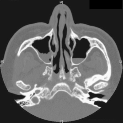
The FP reviewed the CT scan and noted that the patient had air-fluid levels in both maxillary sinuses and loculated fluid on the right side. The patient was given a diagnosis of bilateral maxillary sinusitis that was not responding to the original antibiotic.
The causes of sinusitis include:
○ infection, which is is typically viral (eg, rhinovirus, parainfluenza, influenza), but may be bacterial (eg, Streptococcus pneumoniae, Haemophilus influenzae, Moraxella catarrhalis). In immunocompromised patients, fulminant fungal sinusitis may occur (eg, rhinocerebral mucormycosis).
○ a noninfectious obstruction, which may be the result of allergies, polyposis, barotrauma (eg, deep-sea diving, airplane travel), chemical irritants, tumors, or conditions that alter mucous composition (eg, cystic fibrosis).
Most sinus infections involve the maxillary sinus followed in frequency by the ethmoid (anterior), frontal, and sphenoid sinuses. Of note: Most cases involve more than one sinus. In acute disease, CT scanning is generally reserved for persistent or recurrent symptoms to confirm sinusitis, or to investigate infectious complications. The modest benefit of antibiotics for improving rates of clinical cure or improvement at 7 to 12 days must be weighed against the risks of harm (primarily gastrointestinal, but also skin rash, vaginal discharge, headache, dizziness, and fatigue).
In this case, the FP changed the patient’s antibiotic to amoxicillin/clavulanate and gave her information about nasal saline irrigation for symptom relief. The FP planned to send the patient to an ear, nose, and throat specialist for further evaluation if her symptoms didn’t improve.
Photos courtesy of Chris McMains, MD. Text for Photo Rounds Friday courtesy of Richard P. Usatine, MD. This case was adapted from: Smith M. Sinusitis. In: Usatine R, Smith M, Mayeaux EJ, et al, eds. Color Atlas of Family Medicine. 2nd ed. New York, NY: McGraw-Hill; 2013:197-202.
To learn more about the Color Atlas of Family Medicine, see: http://www.amazon.com/Color-Family-Medicine-Richard-Usatine/dp/0071769641/ref=dp_ob_title_bk
You can now get the second edition of the Color Atlas of Family Medicine as an app for mobile devices by clicking on this link: http://usatinemedia.com/
