User login
- Position yourself so your upper trunk, arm, and hand move as a unit to elevate the head.
- Elevate the head to the level of the uterine incision, rather than bringing the incision down to the head.
- Rotate the occiput anteriorly to present the shortest fetal head diameters to the incision.
- Reduce the lower lip of the uterine incision beneath the fetal head, as you would reduce a posterior cervical lip at a vaginal delivery.
Although it’s a skill vital for cesarean birth, manual delivery of the fetal head from a low pelvic station is given only cursory coverage in standard obstetrics texts and reviews.1-4 To learn successful methods, therefore, physicians are forced to rely on observation, anecdotal experience, and—least desirably—trial and error.
Possibly the most frequently employed maneuver in this scenario is to have an assistant elevate the head with his or her hand.1,3,4 However, this technique may not provide sufficient elevation. Furthermore, subsequent manipulation by the clinician may extend the uterine incision and risk injury to the uterine vessels and bladder. It is an untested assumption that such extensions increase the risk of uterine rupture during subsequent trials of labor.
With the goals of minimizing delay, head compression, and strain on the uterine incision, I developed the elevate, rotate, and reduce (ERR) technique for expeditious delivery of the head from a deep pelvic station.
Approaching the head
Begin the procedure by identifying the fetal position; if it is occiput posterior, you may wish to have a disposable vacuum extractor available. Then stand so that your dominant hand is closest to the patient’s pelvis.
When making the abdominal incision, consider fetal size and maternal body habitus. Be sure all layers of the incision are long enough to permit manipulation and delivery—don’t let surgical pride force you to struggle with an inadequate Pfannenstiel incision. Be prepared to identify and extend any layer of the incision that gives considerable resistance at the time of delivery.
Before proceeding to the uterine incision, make sure there is adequate rectus separation. Check the clearance by separating the right and left bodies of the rectus muscle with manual traction. If more room is needed, create a “partial Maylard incision” by cutting bilateral transverse incisions with Mayo scissors across the medial third of each body. This gives the fetus more room while avoiding damage to the inferior epigastric vessels.
The uterine incision should be as wide as the mean fetal head diameter—usually the same 10 cm that we expect of the fully dilated cervix at term. In patients with an undeveloped lower uterine segment, varices, adhesions, or leiomyomata, there may be insufficient width for a low-transverse incision. In these cases you may need to convert to a U, J, or vertical incision.
Take advantage of a well-developed lower uterine segment and avoid tight U- or V-shaped transverse incisions. Identify the uterine vessels with adequate retraction, and incise laterally right up to them. An alternate technique would be to extend the initial incision by pulling apart the ends with your index fingers, laterally and cephalad (towards the mother’s axillae). Blunt dissection may cause less bleeding from the uterine vessels.5
The ERR technique
“Don’t break the wrist” is a classic admonition, familiar to generations of obstetricians. It warns against a practice that begins by reaching for the head with extended wrist, not elevating the head enough, and delivering the head from whatever position it presents in. The head is delivered with firm wrist flexion (breaking, cocking, or levering the wrist) and anterior traction. This puts caudal leverage upon the lower edge of the uterine incision, which may cause an extension down to the bladder or into the cervix. The ERR technique corrects these errors and minimizes strain on the incision.
Elevate. The first goal of ERR is to elevate the fetal vertex to the lower edge of the uterine incision. Bringing the incision down to the presenting part may strain it. Start by holding the fingers of your dominant hand together, with the thumb tucked against the lateral base Safe delivery of the fetal head during cesarean section of the index finger and the fingers extended straight.
Next, insert your fingertips into the uterine incision and advance them around the fetal head—but not while standing erect with your wrist extended. Such a posture would cause 2 problems: First, you would have insufficient leverage and strength to advance your fingers deep into the pelvis. Second, the angle of your wrist would be obstructed by the uterine incision, the lower edge of the abdominal incision, and the pubis.
Instead, try 1 of the following 2 approaches: The first option is to extend your arm in a straight line from shoulder to fingertip, keeping it almost parallel to the patient’s longitudinal axis. This is accomplished by flexing forward at the waist while facing cross-table, and abducting your arm out from your shoulder. Another option is to turn and face the head of the table, flex at the waist, and extend your arm behind you while pronating your hand. Both of these positions keep your wrist angle neutral and allow you to use the strength and leverage of your entire upper trunk to move your hand.
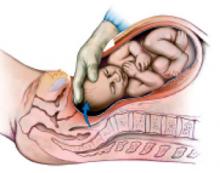
In order to elevate the head, your fingers need to achieve at least a quarter-circle grip around the vertex (FIGURE 1). If the head is so deep or tightly applied that you cannot achieve this in 1 continuous movement, proceed in stages. First, have an assistant apply transvaginal digital pressure to elevate the head from below. Then, advance your fingers as far as you can around the fetal head while flexing and extending the fingers in a worm-like wiggling movement. Flex and lock your fingers against the fetal cranium, and lean your entire upper trunk and arm away from the maternal pelvis. This generally elevates the head by roughly a centimeter, enough to advance your fingers a little further. Repeat these motions until your fingers have achieved the desired quarter-circle, at which point your assistant may withdraw from below.
Now lock your arm from shoulder to fingertip, and lean out of the patient’s pelvis 1 more time. You may note the sensation and sound of breaking suction as the fetal head escapes the pelvic grip. Maintain this steady traction for several seconds. When the vertex has reached the level of the lower margin of the uterine incision, you can rotate the fetus.
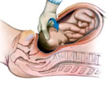
Rotate. Before you begin rotation, confirm the fetal position (the fetal ears are useful as a landmark). If the head is not already in occiput anterior, grasp it and rotate the occiput into the incision (FIGURE 2). As in vaginal delivery, this presents the shortest fetal-head diameters to the birth orifice. If the head has been markedly molded by labor, rotation will bring the narrow end of a dilating cone into the uterine incision. This helps direct the head anteriorly, rather than back into the pelvis, when fundal pressure is applied.
Reduce. Using your hand like a pair of forceps may cause uterine extensions or lacerations if the uterine incision is not large enough to accommodate both your hand and the fetal head. Instead, use 4 fingers in shoehorn fashion to guide the head out of the incision. First, however, there is usually another obstacle to overcome: Fundal pressure may merely force the head back toward the pelvis. Your fingers cannot present a shallow enough angle to direct the head anteriorly, unless your hand is in so deep that your palm fills the uterine incision.
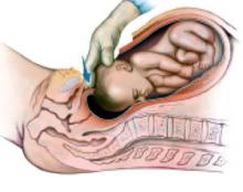
You may substitute instruments for the guiding fingers, using the Murless head extractor, the Torpin vectis blade, or 1 blade of a short-shank Simpson forceps. However, a lack of availability may limit your choices. Although you may use a disposable vacuum extractor system, consider saving that expense with this alternative: Using your fingertips, reduce the lower lip of the uterine incision beneath the fetal head, as you would reduce a posterior cervical lip at a vaginal delivery. (FIGURE 3). The reduced lip tends to extend the head anteriorly and direct it away from the pelvis. Withdraw your hand so only your fingertips remain to guide the head, while the back of your fingers retract the lower abdominal wall. With fundal pressure the head should move anteriorly out of the abdomen.
A head in the occiput-posterior position may resist attempts to rotate to occiput anterior. Should this occur, try following the incision reduction with direct manual flexion of the head out of the abdomen. If reduction flexion puts excess strain on the incision, abandon the attempt. Instead, use a disposable vacuum extractor cup—placed on the fetal brow as close to the vertex as possible—to flex the head out of the incision. Should this fail as well, extend the low-transverse uterine incision cranially into a generous U-incision, and repeat the vacuum procedure.
FIGURE 1 THE ERR SEQUENCE
Elevate. Lock the fingers into a quarter-circle around the vertex. Apply traction out of the pelvis with the hand and the entire extended arm.
FIGURE 2 THE ERR SEQUENCE
Rotate. Grasp the fetal head between the thumb and fingers and rotate it so the occiput faces the incision.
FIGURE 3 THE ERR SEQUENCE
Reduce. Push the lower edge of the uterine incision down until it is posterior to the fetal head.
Conclusion
This technique for delivering the fetal head offers minimal risk of uterine laceration and lends itself to instruction and recollection.
I have not evaluated ERR by clinical trial, both due to absence of a gold-standard technique for the control group and because ERR has yielded satisfactory outcomes in the 20 years over which I have developed, applied, and taught it. Still, the technique raises opportunities for clinical investigation. For example, residency training programs concerned over their incidence of uterine extensions and lacerations may wish to adopt this method and subsequently produce a historical-controlled, retrospective cohort study as a quality-improvement project.
Dr. Chao reports no financial relationship with any companies whose products are mentioned in this article.
1. Cunningham FG, MacDonald PC, Gant NF, et al. Williams Obstetrics. 20th ed. Stamford, Conn: Appleton & Lange; 1997;518.-
2. Scott JR. Cesarean delivery. In: Scott JR, Disaia PJ, Hammond CB, Spellacy WN, eds. Danforth’s Obstetrics and Gynecology. 8th ed. Philadelphia: Lippincott Williams and Wilkins; 1999;462.-
3. Depp R. Cesarean delivery. In: Gabbe SG, Niebyl JR, Simpson JL. Obstetrics: Normal and Problem Pregnancies. 4th ed. New York: Churchill Livingstone, 2002;554.-
4. Field CS. Surgical techniques for cesarean section. Obstet Gynecol Clin NA. 1988;15:664-665.
5. Magann EF, Chauhan SP, Bufkin L, Field K, Roberts WE, Martin JN, Jr. Intraoperative hemorrhage by blunt versus sharp expansion of the uterine incision at caesarean delivery: a randomized clinical trial. BJOG. 2002;109:448-452.
- Position yourself so your upper trunk, arm, and hand move as a unit to elevate the head.
- Elevate the head to the level of the uterine incision, rather than bringing the incision down to the head.
- Rotate the occiput anteriorly to present the shortest fetal head diameters to the incision.
- Reduce the lower lip of the uterine incision beneath the fetal head, as you would reduce a posterior cervical lip at a vaginal delivery.
Although it’s a skill vital for cesarean birth, manual delivery of the fetal head from a low pelvic station is given only cursory coverage in standard obstetrics texts and reviews.1-4 To learn successful methods, therefore, physicians are forced to rely on observation, anecdotal experience, and—least desirably—trial and error.
Possibly the most frequently employed maneuver in this scenario is to have an assistant elevate the head with his or her hand.1,3,4 However, this technique may not provide sufficient elevation. Furthermore, subsequent manipulation by the clinician may extend the uterine incision and risk injury to the uterine vessels and bladder. It is an untested assumption that such extensions increase the risk of uterine rupture during subsequent trials of labor.
With the goals of minimizing delay, head compression, and strain on the uterine incision, I developed the elevate, rotate, and reduce (ERR) technique for expeditious delivery of the head from a deep pelvic station.
Approaching the head
Begin the procedure by identifying the fetal position; if it is occiput posterior, you may wish to have a disposable vacuum extractor available. Then stand so that your dominant hand is closest to the patient’s pelvis.
When making the abdominal incision, consider fetal size and maternal body habitus. Be sure all layers of the incision are long enough to permit manipulation and delivery—don’t let surgical pride force you to struggle with an inadequate Pfannenstiel incision. Be prepared to identify and extend any layer of the incision that gives considerable resistance at the time of delivery.
Before proceeding to the uterine incision, make sure there is adequate rectus separation. Check the clearance by separating the right and left bodies of the rectus muscle with manual traction. If more room is needed, create a “partial Maylard incision” by cutting bilateral transverse incisions with Mayo scissors across the medial third of each body. This gives the fetus more room while avoiding damage to the inferior epigastric vessels.
The uterine incision should be as wide as the mean fetal head diameter—usually the same 10 cm that we expect of the fully dilated cervix at term. In patients with an undeveloped lower uterine segment, varices, adhesions, or leiomyomata, there may be insufficient width for a low-transverse incision. In these cases you may need to convert to a U, J, or vertical incision.
Take advantage of a well-developed lower uterine segment and avoid tight U- or V-shaped transverse incisions. Identify the uterine vessels with adequate retraction, and incise laterally right up to them. An alternate technique would be to extend the initial incision by pulling apart the ends with your index fingers, laterally and cephalad (towards the mother’s axillae). Blunt dissection may cause less bleeding from the uterine vessels.5
The ERR technique
“Don’t break the wrist” is a classic admonition, familiar to generations of obstetricians. It warns against a practice that begins by reaching for the head with extended wrist, not elevating the head enough, and delivering the head from whatever position it presents in. The head is delivered with firm wrist flexion (breaking, cocking, or levering the wrist) and anterior traction. This puts caudal leverage upon the lower edge of the uterine incision, which may cause an extension down to the bladder or into the cervix. The ERR technique corrects these errors and minimizes strain on the incision.
Elevate. The first goal of ERR is to elevate the fetal vertex to the lower edge of the uterine incision. Bringing the incision down to the presenting part may strain it. Start by holding the fingers of your dominant hand together, with the thumb tucked against the lateral base Safe delivery of the fetal head during cesarean section of the index finger and the fingers extended straight.
Next, insert your fingertips into the uterine incision and advance them around the fetal head—but not while standing erect with your wrist extended. Such a posture would cause 2 problems: First, you would have insufficient leverage and strength to advance your fingers deep into the pelvis. Second, the angle of your wrist would be obstructed by the uterine incision, the lower edge of the abdominal incision, and the pubis.
Instead, try 1 of the following 2 approaches: The first option is to extend your arm in a straight line from shoulder to fingertip, keeping it almost parallel to the patient’s longitudinal axis. This is accomplished by flexing forward at the waist while facing cross-table, and abducting your arm out from your shoulder. Another option is to turn and face the head of the table, flex at the waist, and extend your arm behind you while pronating your hand. Both of these positions keep your wrist angle neutral and allow you to use the strength and leverage of your entire upper trunk to move your hand.
In order to elevate the head, your fingers need to achieve at least a quarter-circle grip around the vertex (FIGURE 1). If the head is so deep or tightly applied that you cannot achieve this in 1 continuous movement, proceed in stages. First, have an assistant apply transvaginal digital pressure to elevate the head from below. Then, advance your fingers as far as you can around the fetal head while flexing and extending the fingers in a worm-like wiggling movement. Flex and lock your fingers against the fetal cranium, and lean your entire upper trunk and arm away from the maternal pelvis. This generally elevates the head by roughly a centimeter, enough to advance your fingers a little further. Repeat these motions until your fingers have achieved the desired quarter-circle, at which point your assistant may withdraw from below.
Now lock your arm from shoulder to fingertip, and lean out of the patient’s pelvis 1 more time. You may note the sensation and sound of breaking suction as the fetal head escapes the pelvic grip. Maintain this steady traction for several seconds. When the vertex has reached the level of the lower margin of the uterine incision, you can rotate the fetus.
Rotate. Before you begin rotation, confirm the fetal position (the fetal ears are useful as a landmark). If the head is not already in occiput anterior, grasp it and rotate the occiput into the incision (FIGURE 2). As in vaginal delivery, this presents the shortest fetal-head diameters to the birth orifice. If the head has been markedly molded by labor, rotation will bring the narrow end of a dilating cone into the uterine incision. This helps direct the head anteriorly, rather than back into the pelvis, when fundal pressure is applied.
Reduce. Using your hand like a pair of forceps may cause uterine extensions or lacerations if the uterine incision is not large enough to accommodate both your hand and the fetal head. Instead, use 4 fingers in shoehorn fashion to guide the head out of the incision. First, however, there is usually another obstacle to overcome: Fundal pressure may merely force the head back toward the pelvis. Your fingers cannot present a shallow enough angle to direct the head anteriorly, unless your hand is in so deep that your palm fills the uterine incision.
You may substitute instruments for the guiding fingers, using the Murless head extractor, the Torpin vectis blade, or 1 blade of a short-shank Simpson forceps. However, a lack of availability may limit your choices. Although you may use a disposable vacuum extractor system, consider saving that expense with this alternative: Using your fingertips, reduce the lower lip of the uterine incision beneath the fetal head, as you would reduce a posterior cervical lip at a vaginal delivery. (FIGURE 3). The reduced lip tends to extend the head anteriorly and direct it away from the pelvis. Withdraw your hand so only your fingertips remain to guide the head, while the back of your fingers retract the lower abdominal wall. With fundal pressure the head should move anteriorly out of the abdomen.
A head in the occiput-posterior position may resist attempts to rotate to occiput anterior. Should this occur, try following the incision reduction with direct manual flexion of the head out of the abdomen. If reduction flexion puts excess strain on the incision, abandon the attempt. Instead, use a disposable vacuum extractor cup—placed on the fetal brow as close to the vertex as possible—to flex the head out of the incision. Should this fail as well, extend the low-transverse uterine incision cranially into a generous U-incision, and repeat the vacuum procedure.
FIGURE 1 THE ERR SEQUENCE
Elevate. Lock the fingers into a quarter-circle around the vertex. Apply traction out of the pelvis with the hand and the entire extended arm.
FIGURE 2 THE ERR SEQUENCE
Rotate. Grasp the fetal head between the thumb and fingers and rotate it so the occiput faces the incision.
FIGURE 3 THE ERR SEQUENCE
Reduce. Push the lower edge of the uterine incision down until it is posterior to the fetal head.
Conclusion
This technique for delivering the fetal head offers minimal risk of uterine laceration and lends itself to instruction and recollection.
I have not evaluated ERR by clinical trial, both due to absence of a gold-standard technique for the control group and because ERR has yielded satisfactory outcomes in the 20 years over which I have developed, applied, and taught it. Still, the technique raises opportunities for clinical investigation. For example, residency training programs concerned over their incidence of uterine extensions and lacerations may wish to adopt this method and subsequently produce a historical-controlled, retrospective cohort study as a quality-improvement project.
Dr. Chao reports no financial relationship with any companies whose products are mentioned in this article.
1. Cunningham FG, MacDonald PC, Gant NF, et al. Williams Obstetrics. 20th ed. Stamford, Conn: Appleton & Lange; 1997;518.-
2. Scott JR. Cesarean delivery. In: Scott JR, Disaia PJ, Hammond CB, Spellacy WN, eds. Danforth’s Obstetrics and Gynecology. 8th ed. Philadelphia: Lippincott Williams and Wilkins; 1999;462.-
3. Depp R. Cesarean delivery. In: Gabbe SG, Niebyl JR, Simpson JL. Obstetrics: Normal and Problem Pregnancies. 4th ed. New York: Churchill Livingstone, 2002;554.-
4. Field CS. Surgical techniques for cesarean section. Obstet Gynecol Clin NA. 1988;15:664-665.
5. Magann EF, Chauhan SP, Bufkin L, Field K, Roberts WE, Martin JN, Jr. Intraoperative hemorrhage by blunt versus sharp expansion of the uterine incision at caesarean delivery: a randomized clinical trial. BJOG. 2002;109:448-452.


