User login
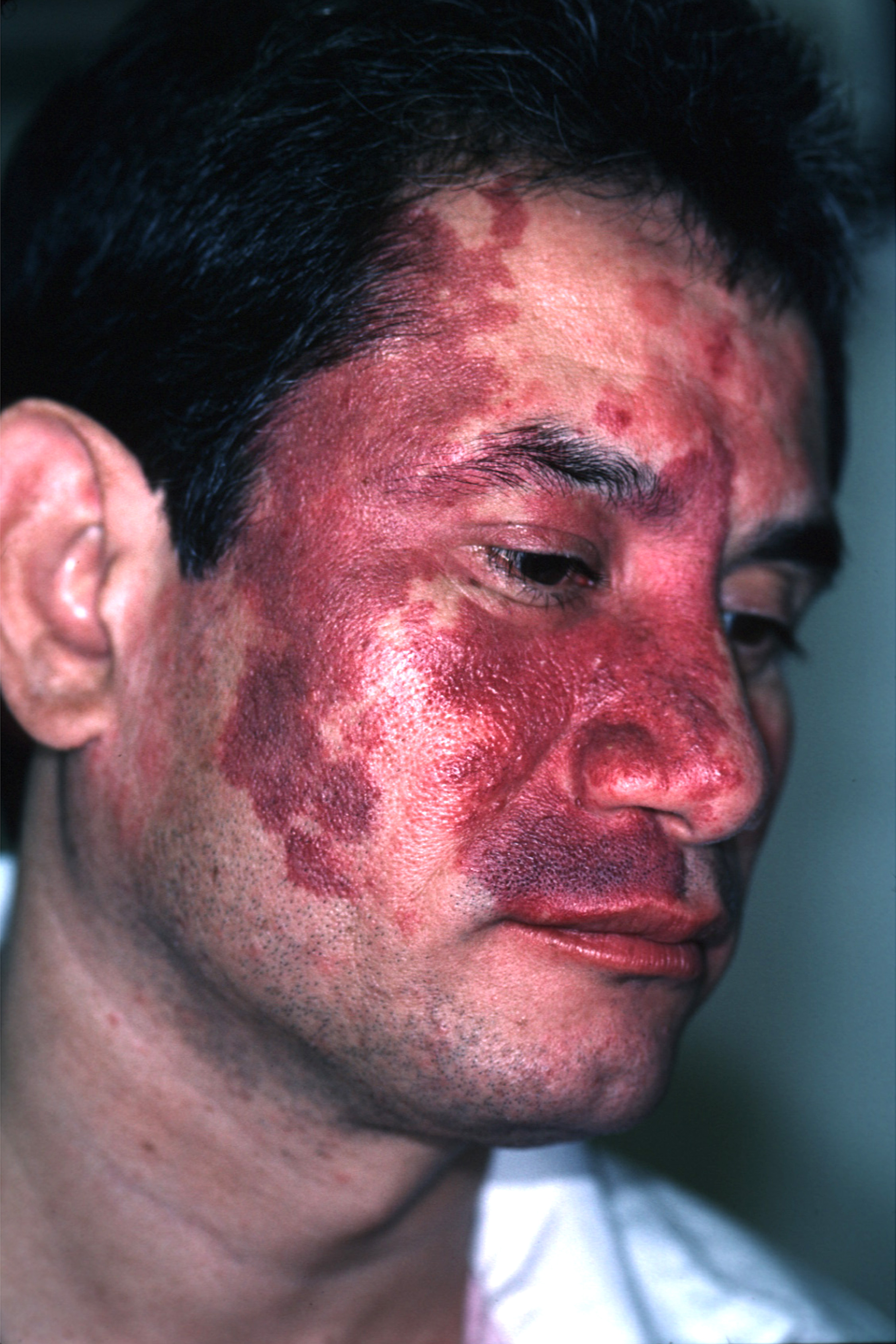
The family physician diagnosed a port-wine stain on the face.
Nevus flammeus, or port-wine stains, are congenital vascular malformations that occur in 0.1 to 0.3% of infants as developmental anomalies. They may be associated with rare syndromes such as Klippel-Trenaunay syndrome and Sturge-Weber syndrome. Port-wine stains are vascular ectasias or dilatations thought to arise from a deficiency of sympathetic nervous innervation to the blood vessels. Dilated capillaries are present throughout the dermis layer of the skin.
Port-wine stains are irregular red-to-purple patches that start out smooth in infancy but may develop hypertrophy and a cobblestone texture with age. Nuchal port-wine stains are associated with alopecia areata. Klippel-Trenaunay syndrome is characterized by vascular malformations, venous varicosities, and soft tissue hyperplasia. Patients with Sturge-Weber syndrome often have mental retardation, epilepsy, and eye problems.
Port-wine stains tend to affect the face and neck, although lesions may affect any body surface, including mucous membranes. Lesions of Klippel-Trenaunay syndrome tend to affect the lower extremities. A diagnosis of Sturge-Weber syndrome requires that a port-wine stain be present in the V1 trigeminal nerve distribution.
Patients with port-wine stains of the eyelids, bilateral trigeminal lesions, and unilateral lesions involving all 3 divisions of the trigeminal nerve are particularly at risk for Sturge-Weber syndrome. The FP noted that this patient had V1 and V2 involvement along with the eyelid.
If Sturge-Weber syndrome is suspected, perform neuroimaging and glaucoma testing. Neuroimaging may reveal leptomeningeal malformations ipsilateral to the port-wine stain. An electroencephalogram may reveal epilepsy. Elevated ocular pressures or visual field deficits may indicate glaucoma. The patient described here denied epilepsy and eye problems and showed no signs of mental retardation. The physician suggested a screening eye exam just to be sure.
Port-wine stains may be treated with make-up. Pulsed-dye laser treatment is another option, albeit an expensive one. Laser treatments blanch most port-wine lesions to some degree, but complete resolution is difficult to achieve and the recurrence rate is high. Patients with port-wine stains should have periodic skin checks as other lesions may develop within the port-wine stains.
After discussing the port-wine stain with the patient, the physician went on to diagnose and treat his cough.
Photos and text for Photo Rounds Friday courtesy of Richard P. Usatine, MD. This case was adapted from: Hitzeman N. Hereditary vascular lesions in adults. In: Usatine R, Smith M, Mayeaux EJ, et al, eds. The Color Atlas of Family Medicine. New York, NY: McGraw-Hill; 2009:865-868.
To learn more about The Color Atlas of Family Medicine, see:
• http://www.amazon.com/Color-Atlas-Family-Medicine/dp/0071474641
You can now get The Color Atlas of Family Medicine as an app for mobile devices including the iPhone and iPad by clicking this link: