User login
We have long understood that vasculitic conditions have various clinical manifestations. The Chapel Hill Consensus Conference classification of systemic vasculitis in 19941 contributed significantly to our understanding of the spectrum of vasculitides and their manifestations, enhancing our diagnostic ability and the likelihood of appropriate treatment.
The ophthalmic manifestations of vasculitis are protean and nonspecific, and should be considered in the overall context of the disease. Patients should be evaluated with the following questions in mind:
- Are the manifestations related to the vasculitis itself?
- Are the manifestations a result or complication of therapy?
- Are the manifestations signs of a completely unrelated and superimposed condition?
This article reviews the three areas of ocular inflammation related to vasculitis and comments on the role of tissue biopsy in the management of these patients.
THREE AREAS OF OCULAR INFLAMMATION
Orbital inflammation
Orbital disease can affect the lacrimal gland (inflammatory dacryoadenitis), extraocular muscles (orbital myositis), and the orbital soft tissues (inflammatory orbital pseudotumor). Orbital inflammation is characterized by relatively sudden onset (within days) of pain, erythema, and proptosis. Diplopia and visual loss from either compression or inflammation of the optic nerve or nerve sheath may be present. Depending upon the structures involved and the degree of involvement, orbital inflammation can be sight-threatening.
Either computed tomography or magnetic resonance imaging should be performed to assess orbital or extraorbital involvement. The orbital structures are particularly amenable to biopsy, which, in this author’s opinion, should be performed whenever possible. The biopsy may need to be interpreted within the context of previous or concurrent immunosuppressive therapy, which can alter the histologic picture, minimize inflammation, and make detection of vasculitis difficult. In addition to identifying inflammation, biopsy helps to identify fungal infection or lymphoma that can follow prolonged immunosuppressive therapy.
Treatment of orbital inflammation requires corticosteroid therapy or some other type of systemic immunosuppression.
Ocular, or globe, inflammation
Episcleritis: observation or topical therapy. Episcleritis usually manifests as an otherwise asymptomatic red eye with typical sector-shaped inflammation. Pain is generally not an issue, although patients often report that the eye does not feel normal. Vision is unaffected and there is no potential threat to sight.
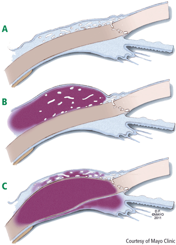
The slit-lamp examination shows dilated vessels in the episcleral tissues that blanch after instillation of a drop of 10% phenylephrine. Simple observation may be the best management course, but topical nonsteroidal anti-inflammatory drugs (NSAIDs) or topical corticosteroids may help some patients who have discomfort. There is probably a spectrum of disease in that some patients may have either severe episcleritis or mild scleritis (Figure 1B). At times it can be difficult to differentiate between severe episcleritis and mild scleritis. Although scleritis generally requires systemic therapy, topical therapy is justified for mild scleritis. Episcleritis is associated with systemic disease in approximately 36% of patients.2–4
Scleritis: may be sight-threatening; requires systemic therapy. Scleritis characteristically presents with intense pain and a red eye.3,5–7 Patients may be sensitive to light and their vision may be compromised. Cataracts and glaucoma can complicate the course of scleritis.
With slit-lamp examination, the redness does not blanch upon instillation of topical 10% phenylephrine as it does with episcleritis. The adjacent cornea may also be affected (Figure 1C). Healed scleritis leaves an area of thinned sclera that appears as a visible blue spot, so if the patient’s history includes red eye with pain and a blue area is visible, the clinician can be confident that a prior episode of scleritis occurred.
Scleritis can be anterior or posterior, and the implications are slightly different for each type. Anterior scleritis can be subclassified as diffuse, nodular, or necrotizing. The necrotizing type can be characterized by painful inflammation or, in the case of scleromalacia perforans, no inflammation and no pain. Posterior scleritis may have minimal pain.
Akpek et al5 reported on a group of 243 patients with scleritis (average age, 52 years; range, 5 to 93 years) who were followed for an average of 1.7 years (range, 0 to 16.6 years). An associated medical condition was present in 107 (44%) patients. Rheumatologic conditions accounted for 37%, with rheumatoid arthritis being most common; infectious disease, with herpes zoster ophthalmicus being most common, accounted for 7%. Of those with an associated medical condition, 78% had been diagnosed previously; the remaining 22% were diagnosed at presentation or the condition developed during follow-up.
Treatment typically requires systemic therapy with NSAIDs, but more often oral or intravenous corticosteroids or even methotrexate, mycophenolate mofetil, cyclophosphamide, or rituximab may be required. Patients with antineutrophil cytoplasmic antibody (ANCA)–positive disease may require more intensive therapy than those with ANCA-negative disease.
Keratitis: may be sight-threatening. Patients with keratitis should be evaluated in the same spirit as patients with scleritis (Figure 1C). Although many patients may have superficial keratitis, which is often related to a dry eye and has no prognostic significance, deep or peripheral ulcerative keratitis is not only consistent with systemic vasculitis but also sight-threatening. Symptoms similar to those observed with scleritis typically include severe pain and photophobia and, as with scleritis, treatment usually involves systemic therapy.
Intraocular inflammation
There is no specific treatment for the eye other than treating the underlying condition. Vascular occlusions can sometimes give rise to neovascularization and patients should be followed for this possibility. As with a central nervous system ischemic event, recovery can be variable.
Uveitis. The term “uvea,” derived from the Greek word for grape, describes the shape of the iris, ciliary body, and choroid. Uveitis is a generic term for intraocular inflammation affecting any or all of these structures.
Iritis, or anterior uveitis, is a frequent accompaniment of keratitis or scleritis. Primarily uveitic involvement with retinal vessel vasculitis involving both arteries and veins is uncommon in general but typical of Behçet disease, especially if a hypopyon uveitis is present.
Anterior uveitis can be treated with topical corticosteroids and cycloplegic drugs, but middle and posterior uveitis almost always requires systemic therapy. Most recently, use of anti–tumor necrosis factor-α drugs has been effective in treating Behçet uveitis.8 The visual prognosis with Behçet disease remains guarded.
GRANULOMATOSIS WITH POLYANGIITIS: EYE INVOLVEMENT IS COMMON
In terms of specific small-vessel vasculitic diseases that affect the eye, granulomatosis with polyangiitis (GPA [Wegener’s granulomatosis]) is the quintessential condition. In data obtained from the Wegener Granulomatosis Support Group,9 eye involvement was noted at presentation in 211 of 701 patients (30%), and during the course of their disease an additional 147 patients developed eye involvement. From the time of initial presentation through the course of follow-up, 359 of the 701 patients (51%) eventually had some type of eye involvement.
In a series of patients seen at the Mayo Clinic,10 orbital inflammatory disease and scleritis were the two most frequent manifestations of eye involvement with GPA. Orbital involvement typically presents with pain, erythema, swelling, and proptosis. Varying degrees of ptosis, diplopia, or visual loss may also be present. Imaging may show an infiltrate that is usually adjacent to the maxillary or ethmoid sinus. This same process can affect the superior temporal orbital quadrant, an area apart from any sinus, and involve the lacrimal gland.
BIOPSY IS ADVISED
Biopsy, either incisional, at times to include debulking, or excisional if possible, is recommended to establish a diagnosis or aid in the selection of therapy. Orbital disease has been observed to progress in patients who are receiving maintenance therapy with methotrexate and have no evidence of systemic disease activity. Acute and chronic inflammation with evidence of active vasculitis is usually seen histologically. Personal observations suggest that intraorbital corticosteroid injection followed by rituximab has been effective therapy for this limited subset of patients. Diagnostic biopsies often must be interpreted in light of partial treatment, making histopathologic diagnosis challenging at times. Biopsy is important for exclusion of lymphoproliferative disease or fungal infection.
CONCLUSION
Underlying vasculitis might play a role in patients with nonspecific ocular presentations. It is essential that the ophthalmologist collaborate with a specialist in vasculitis (and vice versa) for evaluation and subsequent therapy, which often involves some form of immunosuppression.
- Jennette JC, Falk RJ, Andrassy K, et al. Nomenclature of systemic vasculitides: proposal of an International Consensus Conference. Arthritis Rheum 1994; 37:187–192.
- Pavesio CE, Meier FM. Systemic disorders associated with episcleritis and scleritis. Curr Opin Ophthalmol 2001; 12:471–478.
- Jabs DA, Mudun A, Dunn JP, Marsh MJ. Episcleritis and scleritis: clinical features and treatment results. Am J Ophthalmol 2000; 130:469–476.
- Akpek EK, Uy HS, Christen W, Gurdal C, Foster CS. Severity of episcleritis and systemic disease association. Ophthalmology 1999; 106:729–731.
- Akpek EK, Thorne JE, Qazi FA, Do DV, Jabs DA. Evaluation of patients with scleritis for systemic disease. Ophthalmology 2004; 111:501–506.
- McCluskey PJ, Watson PG, Lightman S, Haybittle J, Restori M, Branley M. Posterior scleritis: clinical features, systemic associations, and outcome in a large series of patients. Ophthalmology 1999; 106:2380–2386.
- Riono WP, Hidayat AA, Rao NA. Scleritis: a clinicopathologic study of 55 cases. Ophthalmology 1999; 106:1328–1333.
- Tabbara KF, Al-Hemidan AI. Infliximab effects compared to conventional therapy in the management of retinal vasculitis in Behçet disease [published online ahead of print October 17, 2008]. Am J Ophthalmol 2008; 146:845–850. doi: 10.1016/j.ajo.2008.09.010
- Abdou NI, Kullman GJ, Hoffman GS, et al. Wegener’s granulomatosis— survey of 701 patients in North America: changes in outcome in the 1990s. J Rheumatol 2002; 29:309–316.
- Bullen CL, Liesegang TJ, McDonald TJ, DeRemee RA. Ocular complications of Wegener’s granulomatosis. Ophthalmology 1983; 90:279–290.
We have long understood that vasculitic conditions have various clinical manifestations. The Chapel Hill Consensus Conference classification of systemic vasculitis in 19941 contributed significantly to our understanding of the spectrum of vasculitides and their manifestations, enhancing our diagnostic ability and the likelihood of appropriate treatment.
The ophthalmic manifestations of vasculitis are protean and nonspecific, and should be considered in the overall context of the disease. Patients should be evaluated with the following questions in mind:
- Are the manifestations related to the vasculitis itself?
- Are the manifestations a result or complication of therapy?
- Are the manifestations signs of a completely unrelated and superimposed condition?
This article reviews the three areas of ocular inflammation related to vasculitis and comments on the role of tissue biopsy in the management of these patients.
THREE AREAS OF OCULAR INFLAMMATION
Orbital inflammation
Orbital disease can affect the lacrimal gland (inflammatory dacryoadenitis), extraocular muscles (orbital myositis), and the orbital soft tissues (inflammatory orbital pseudotumor). Orbital inflammation is characterized by relatively sudden onset (within days) of pain, erythema, and proptosis. Diplopia and visual loss from either compression or inflammation of the optic nerve or nerve sheath may be present. Depending upon the structures involved and the degree of involvement, orbital inflammation can be sight-threatening.
Either computed tomography or magnetic resonance imaging should be performed to assess orbital or extraorbital involvement. The orbital structures are particularly amenable to biopsy, which, in this author’s opinion, should be performed whenever possible. The biopsy may need to be interpreted within the context of previous or concurrent immunosuppressive therapy, which can alter the histologic picture, minimize inflammation, and make detection of vasculitis difficult. In addition to identifying inflammation, biopsy helps to identify fungal infection or lymphoma that can follow prolonged immunosuppressive therapy.
Treatment of orbital inflammation requires corticosteroid therapy or some other type of systemic immunosuppression.
Ocular, or globe, inflammation
Episcleritis: observation or topical therapy. Episcleritis usually manifests as an otherwise asymptomatic red eye with typical sector-shaped inflammation. Pain is generally not an issue, although patients often report that the eye does not feel normal. Vision is unaffected and there is no potential threat to sight.
The slit-lamp examination shows dilated vessels in the episcleral tissues that blanch after instillation of a drop of 10% phenylephrine. Simple observation may be the best management course, but topical nonsteroidal anti-inflammatory drugs (NSAIDs) or topical corticosteroids may help some patients who have discomfort. There is probably a spectrum of disease in that some patients may have either severe episcleritis or mild scleritis (Figure 1B). At times it can be difficult to differentiate between severe episcleritis and mild scleritis. Although scleritis generally requires systemic therapy, topical therapy is justified for mild scleritis. Episcleritis is associated with systemic disease in approximately 36% of patients.2–4
Scleritis: may be sight-threatening; requires systemic therapy. Scleritis characteristically presents with intense pain and a red eye.3,5–7 Patients may be sensitive to light and their vision may be compromised. Cataracts and glaucoma can complicate the course of scleritis.
With slit-lamp examination, the redness does not blanch upon instillation of topical 10% phenylephrine as it does with episcleritis. The adjacent cornea may also be affected (Figure 1C). Healed scleritis leaves an area of thinned sclera that appears as a visible blue spot, so if the patient’s history includes red eye with pain and a blue area is visible, the clinician can be confident that a prior episode of scleritis occurred.
Scleritis can be anterior or posterior, and the implications are slightly different for each type. Anterior scleritis can be subclassified as diffuse, nodular, or necrotizing. The necrotizing type can be characterized by painful inflammation or, in the case of scleromalacia perforans, no inflammation and no pain. Posterior scleritis may have minimal pain.
Akpek et al5 reported on a group of 243 patients with scleritis (average age, 52 years; range, 5 to 93 years) who were followed for an average of 1.7 years (range, 0 to 16.6 years). An associated medical condition was present in 107 (44%) patients. Rheumatologic conditions accounted for 37%, with rheumatoid arthritis being most common; infectious disease, with herpes zoster ophthalmicus being most common, accounted for 7%. Of those with an associated medical condition, 78% had been diagnosed previously; the remaining 22% were diagnosed at presentation or the condition developed during follow-up.
Treatment typically requires systemic therapy with NSAIDs, but more often oral or intravenous corticosteroids or even methotrexate, mycophenolate mofetil, cyclophosphamide, or rituximab may be required. Patients with antineutrophil cytoplasmic antibody (ANCA)–positive disease may require more intensive therapy than those with ANCA-negative disease.
Keratitis: may be sight-threatening. Patients with keratitis should be evaluated in the same spirit as patients with scleritis (Figure 1C). Although many patients may have superficial keratitis, which is often related to a dry eye and has no prognostic significance, deep or peripheral ulcerative keratitis is not only consistent with systemic vasculitis but also sight-threatening. Symptoms similar to those observed with scleritis typically include severe pain and photophobia and, as with scleritis, treatment usually involves systemic therapy.
Intraocular inflammation
There is no specific treatment for the eye other than treating the underlying condition. Vascular occlusions can sometimes give rise to neovascularization and patients should be followed for this possibility. As with a central nervous system ischemic event, recovery can be variable.
Uveitis. The term “uvea,” derived from the Greek word for grape, describes the shape of the iris, ciliary body, and choroid. Uveitis is a generic term for intraocular inflammation affecting any or all of these structures.
Iritis, or anterior uveitis, is a frequent accompaniment of keratitis or scleritis. Primarily uveitic involvement with retinal vessel vasculitis involving both arteries and veins is uncommon in general but typical of Behçet disease, especially if a hypopyon uveitis is present.
Anterior uveitis can be treated with topical corticosteroids and cycloplegic drugs, but middle and posterior uveitis almost always requires systemic therapy. Most recently, use of anti–tumor necrosis factor-α drugs has been effective in treating Behçet uveitis.8 The visual prognosis with Behçet disease remains guarded.
GRANULOMATOSIS WITH POLYANGIITIS: EYE INVOLVEMENT IS COMMON
In terms of specific small-vessel vasculitic diseases that affect the eye, granulomatosis with polyangiitis (GPA [Wegener’s granulomatosis]) is the quintessential condition. In data obtained from the Wegener Granulomatosis Support Group,9 eye involvement was noted at presentation in 211 of 701 patients (30%), and during the course of their disease an additional 147 patients developed eye involvement. From the time of initial presentation through the course of follow-up, 359 of the 701 patients (51%) eventually had some type of eye involvement.
In a series of patients seen at the Mayo Clinic,10 orbital inflammatory disease and scleritis were the two most frequent manifestations of eye involvement with GPA. Orbital involvement typically presents with pain, erythema, swelling, and proptosis. Varying degrees of ptosis, diplopia, or visual loss may also be present. Imaging may show an infiltrate that is usually adjacent to the maxillary or ethmoid sinus. This same process can affect the superior temporal orbital quadrant, an area apart from any sinus, and involve the lacrimal gland.
BIOPSY IS ADVISED
Biopsy, either incisional, at times to include debulking, or excisional if possible, is recommended to establish a diagnosis or aid in the selection of therapy. Orbital disease has been observed to progress in patients who are receiving maintenance therapy with methotrexate and have no evidence of systemic disease activity. Acute and chronic inflammation with evidence of active vasculitis is usually seen histologically. Personal observations suggest that intraorbital corticosteroid injection followed by rituximab has been effective therapy for this limited subset of patients. Diagnostic biopsies often must be interpreted in light of partial treatment, making histopathologic diagnosis challenging at times. Biopsy is important for exclusion of lymphoproliferative disease or fungal infection.
CONCLUSION
Underlying vasculitis might play a role in patients with nonspecific ocular presentations. It is essential that the ophthalmologist collaborate with a specialist in vasculitis (and vice versa) for evaluation and subsequent therapy, which often involves some form of immunosuppression.
- Jennette JC, Falk RJ, Andrassy K, et al. Nomenclature of systemic vasculitides: proposal of an International Consensus Conference. Arthritis Rheum 1994; 37:187–192.
- Pavesio CE, Meier FM. Systemic disorders associated with episcleritis and scleritis. Curr Opin Ophthalmol 2001; 12:471–478.
- Jabs DA, Mudun A, Dunn JP, Marsh MJ. Episcleritis and scleritis: clinical features and treatment results. Am J Ophthalmol 2000; 130:469–476.
- Akpek EK, Uy HS, Christen W, Gurdal C, Foster CS. Severity of episcleritis and systemic disease association. Ophthalmology 1999; 106:729–731.
- Akpek EK, Thorne JE, Qazi FA, Do DV, Jabs DA. Evaluation of patients with scleritis for systemic disease. Ophthalmology 2004; 111:501–506.
- McCluskey PJ, Watson PG, Lightman S, Haybittle J, Restori M, Branley M. Posterior scleritis: clinical features, systemic associations, and outcome in a large series of patients. Ophthalmology 1999; 106:2380–2386.
- Riono WP, Hidayat AA, Rao NA. Scleritis: a clinicopathologic study of 55 cases. Ophthalmology 1999; 106:1328–1333.
- Tabbara KF, Al-Hemidan AI. Infliximab effects compared to conventional therapy in the management of retinal vasculitis in Behçet disease [published online ahead of print October 17, 2008]. Am J Ophthalmol 2008; 146:845–850. doi: 10.1016/j.ajo.2008.09.010
- Abdou NI, Kullman GJ, Hoffman GS, et al. Wegener’s granulomatosis— survey of 701 patients in North America: changes in outcome in the 1990s. J Rheumatol 2002; 29:309–316.
- Bullen CL, Liesegang TJ, McDonald TJ, DeRemee RA. Ocular complications of Wegener’s granulomatosis. Ophthalmology 1983; 90:279–290.