User login
DURING A ROUTINE EXAM, a 45-year-old Caucasian woman complained of intense itching on her labia. She said that the itching had been an issue for more than 9 months and that she found herself scratching several times a day. She denied any vaginal discharge and said she hadn’t been sexually active in years. She had tried over-the-counter antifungals and topical Benadryl, but they provided only limited relief.
The patient had red thickened plaques with accentuated skin lines (furrows) covering both of her labia majora (FIGURE). Throughout the lesion, there were scattered areas of excoriation. Her labia minor were spared.
FIGURE
Red thickened plaques on labia majora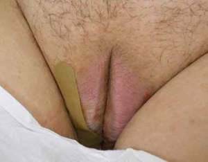
A speculum and bimanual exam were normal. No inguinal lymphadenopathy was present.
WHAT IS YOUR DIAGNOSIS?
HOW WOULD YOU TREAT THIS PATIENT?
Diagnosis: Lichen simplex chronicus
This patient was given a diagnosis of lichen simplex chronicus (circumscribed neurodermatitis)—pathohistologic changes to the skin caused by habitual trauma from scratching a single area. The lesion begins as small red papules that later coalesce into a plaque with furrows (lichenification).1 The plaques become well circumscribed with attenuated skin lines. This is due to the thickening of both the epidermis (acanthosis) and stratum corneum (hyperkeratosis).2 Hyperpigmentation or hypopigmentation may be present—particularly in patients with naturally dark-colored skin.1
The epidemiology of lichen simplex chronicus is unknown. It tends to occur in adults between 30 and 50 years of age (although it has been seen in children) and is more common in women and people of Asian descent.3 The areas of the body that are affected are those that are easily reached, including the:4,5
- outer portion of the lower legs
- scrotum and vulva; pubic and anal areas
- wrists and ankles
- upper eyelids
- back or side of neck
- ear canal
- extensor forearms near the elbow
- fold behind the ear
- scalp
Pathology will show lichenification, acanthosis, hyperkeratosis, and eczematous inflammation resembling psoriasis. The histologic separation between lichen simplex chronicus and psoriasis is particularly difficult, which leads some pathologists to report the process as “psoriasiform dermatitis.”1 That was the case in this presentation.
Differential diagnosis includes lichen planus, psoriasis
Skin conditions with a similar appearance to lichen simplex chronicus include lichen planus, lichen sclerosus, psoriasis, mycosis fungoides, and extramammary Paget’s disease.1,2,4,5
Lichen planus is an inflammatory cutaneous lesion of unknown etiology. It can be found throughout the body, including the mucous membranes. It commonly presents as the 5 Ps: pruritic, planar, polyangular, purple, and papules.5 Upon close examination, Wickham’s striae (reticular white lines) may be visible.4,5
Lichen sclerosus is a cutaneous disease of unknown origin that tends to occur in postmenopausal women; it prompts complaints of pruritus and dyspareunia.5 It presents as white atrophic plaques that may encompass both the vagina and rectum.4,5
Psoriasis lesions are usually distinctive, with red scaling papules that tend to coalesce into plaques. These lesions are associated with Auspitz sign—pinpoint bleeding following removal of silvery white scale.4,5 Lesions are often found on the elbows, groin, knees, scalp, gluteal cleft, fingernails, and toes.4
Mycosis fungoides (MF) is a cutaneous T-cell lymphoma that can have similar clinical characteristics to lichen simplex chronicus. It evolves through 4 phases: pre-MF, patch, plaque, and tumor.4 The patch phase may be confused with lichen simplex chronicus because there are flat, erythematous, and pruritic lesions. It also presents with lymphadenopathy and lesions that are persistently resistant to topical steroid treatments. High clinical suspicion and multiple biopsies at different sites may be useful.
Extramammary Paget’s disease is a rare cutaneous form of adenocarcinoma. About 12% of patients have a concurrent underlying internal malignancy.4,5 It appears as a white-to-red, scaling or macerated, infiltrated, eroded, or ulcerated plaque, most frequently observed on the labia majora and scrotum.5
2 keys to diagnosis
A history of severe pruritus (with a chronic itch-scratch cycle) combined with the findings of lichenification should make you suspect lichen simplex chronicus. It may be necessary to first treat the itch-scratch cycle before you can identify the underlying disease.1 If clinical diagnosis is still unclear, you may need to do a skin biopsy for pathologic identification and to rule out neoplasia.
Look for concomitant psychiatric disorders that often have contributing factors, such as depression, anxiety, and obsessive-compulsive disorder.1,4,5 Correlation of history, physical exam, and pathophysiology is enough for the diagnosis.
Focus Tx on interrupting the itch-scratch cycle
Tell patients with lichen simplex chronicus that a permanent cure may not be possible. Intermittent therapy may be necessary for years.1 The key to long-term success is disruption of the itch-scratch cycle. Often, the scratching occurs during sleep without the patient even being aware. So it’s helpful to educate patients about the itch-scratch cycle and to advise them to keep their nails trimmed, wear gloves at nighttime, and if needed, apply an occlusive dressing.1,2 It’s also helpful to avoid irritants such as harsh soaps and washcloths, panty liners, tight clothing, perfumes, and deodorants.2
Help the patient break the nighttime itch-scratch cycle by prescribing sedating H1 antihistamines such as hydroxyzine (10-25 mg taken within 2 hours of bed time); increase the dose every 7 days until scratching ceases or adverse effects develop.2 Another option is a tricyclic antidepressant—doxepin 25 to 75 mg or amitriptyline 25 to 75 mg— taken within 2 hours of bed time.1
Recommend that patients use lubricants and petroleum-based ointments to restore the damaged skin barrier and its natural function. Using these products after bathing may be especially helpful.1
Class I and II topical steroids can be used as first-line treatment to reduce inflammation and pruritus. Be sure to advise patients of potential adverse effects of potent topical steroids, such as atrophy, discoloration, and striae.1,2,5
Second-line topical treatments include tacrolimus ointment 0.1%, pimecrolimus cream 1%, topical lidocaine 2%, and capsaicin 0.025% to 0.075% cream applied 3 times a day.2 Depending on the size and shape of the lesion, intralesional steroids such as triamcinolone acetate in varying concentrations may be beneficial.
Selective serotonin reuptake inhibitors have been shown to benefit patients during the day, and to address other psychological comorbidities that may be present (eg, anxiety, depression, or obsessive-compulsive disorder).1
Be sure to screen for secondary super-infections (bacterial and fungal) and treat accordingly. Follow-up should be scheduled for 4 weeks after treatment has begun.2
Steroids provided relief for my patient
I prescribed clobetasol 0.05% ointment without occlusion twice a day for 4 weeks. I also prescribed hydroxyzine 25 mg to be taken at bedtime and fluoxetine 20 mg daily for underlying depression.
At follow-up 4 weeks later, my patient reported excellent relief from her pruritus. Her labia’s erythema had greatly decreased, but chronic skin changes were still present. I advised her to apply the clobetasol every 2 weeks as needed, with follow-up in 3 months.
CORRESPONDENCE
Stephen Colden Cahill, DO, Assistant Clinical Professor, Michigan State University, 8300 Westpark Way, Zeeland, MI 49464; [email protected]
1. Stewart KM. Clinical care of vulvar pruritus with emphasis on one common cause, lichen simplex chronicus. Dermatol Clin. 2010;28:669-680.
2. Lynch PJ. Lichen simplex chronicus (atopic/neurodermatitis) of the anogenital region. Dermatol Ther. 2004;17:8-19.
3. Prajapati V, Barankin B. Dermacase. Lichen simplex chronicus. Can Fam Physician. 2008;54:1391-1393.
4. Habif T. Clinical Dermatology: A Color Guide to Diagnosis and Therapy. 4th ed. Philadelphia Pa: Mosby; 2003.
5. Burgin S. Nummular eczema and lichen simplex chronicus/prurigo nodularis. In: Wolff K Goldsmith LA, Katz SI, et al, eds. Fitzpatrick’s Dermatology in General Medicine. 4th ed. New York, NY: McGraw-Hill Companies; 2008: 158–162.
DURING A ROUTINE EXAM, a 45-year-old Caucasian woman complained of intense itching on her labia. She said that the itching had been an issue for more than 9 months and that she found herself scratching several times a day. She denied any vaginal discharge and said she hadn’t been sexually active in years. She had tried over-the-counter antifungals and topical Benadryl, but they provided only limited relief.
The patient had red thickened plaques with accentuated skin lines (furrows) covering both of her labia majora (FIGURE). Throughout the lesion, there were scattered areas of excoriation. Her labia minor were spared.
FIGURE
Red thickened plaques on labia majora
A speculum and bimanual exam were normal. No inguinal lymphadenopathy was present.
WHAT IS YOUR DIAGNOSIS?
HOW WOULD YOU TREAT THIS PATIENT?
Diagnosis: Lichen simplex chronicus
This patient was given a diagnosis of lichen simplex chronicus (circumscribed neurodermatitis)—pathohistologic changes to the skin caused by habitual trauma from scratching a single area. The lesion begins as small red papules that later coalesce into a plaque with furrows (lichenification).1 The plaques become well circumscribed with attenuated skin lines. This is due to the thickening of both the epidermis (acanthosis) and stratum corneum (hyperkeratosis).2 Hyperpigmentation or hypopigmentation may be present—particularly in patients with naturally dark-colored skin.1
The epidemiology of lichen simplex chronicus is unknown. It tends to occur in adults between 30 and 50 years of age (although it has been seen in children) and is more common in women and people of Asian descent.3 The areas of the body that are affected are those that are easily reached, including the:4,5
- outer portion of the lower legs
- scrotum and vulva; pubic and anal areas
- wrists and ankles
- upper eyelids
- back or side of neck
- ear canal
- extensor forearms near the elbow
- fold behind the ear
- scalp
Pathology will show lichenification, acanthosis, hyperkeratosis, and eczematous inflammation resembling psoriasis. The histologic separation between lichen simplex chronicus and psoriasis is particularly difficult, which leads some pathologists to report the process as “psoriasiform dermatitis.”1 That was the case in this presentation.
Differential diagnosis includes lichen planus, psoriasis
Skin conditions with a similar appearance to lichen simplex chronicus include lichen planus, lichen sclerosus, psoriasis, mycosis fungoides, and extramammary Paget’s disease.1,2,4,5
Lichen planus is an inflammatory cutaneous lesion of unknown etiology. It can be found throughout the body, including the mucous membranes. It commonly presents as the 5 Ps: pruritic, planar, polyangular, purple, and papules.5 Upon close examination, Wickham’s striae (reticular white lines) may be visible.4,5
Lichen sclerosus is a cutaneous disease of unknown origin that tends to occur in postmenopausal women; it prompts complaints of pruritus and dyspareunia.5 It presents as white atrophic plaques that may encompass both the vagina and rectum.4,5
Psoriasis lesions are usually distinctive, with red scaling papules that tend to coalesce into plaques. These lesions are associated with Auspitz sign—pinpoint bleeding following removal of silvery white scale.4,5 Lesions are often found on the elbows, groin, knees, scalp, gluteal cleft, fingernails, and toes.4
Mycosis fungoides (MF) is a cutaneous T-cell lymphoma that can have similar clinical characteristics to lichen simplex chronicus. It evolves through 4 phases: pre-MF, patch, plaque, and tumor.4 The patch phase may be confused with lichen simplex chronicus because there are flat, erythematous, and pruritic lesions. It also presents with lymphadenopathy and lesions that are persistently resistant to topical steroid treatments. High clinical suspicion and multiple biopsies at different sites may be useful.
Extramammary Paget’s disease is a rare cutaneous form of adenocarcinoma. About 12% of patients have a concurrent underlying internal malignancy.4,5 It appears as a white-to-red, scaling or macerated, infiltrated, eroded, or ulcerated plaque, most frequently observed on the labia majora and scrotum.5
2 keys to diagnosis
A history of severe pruritus (with a chronic itch-scratch cycle) combined with the findings of lichenification should make you suspect lichen simplex chronicus. It may be necessary to first treat the itch-scratch cycle before you can identify the underlying disease.1 If clinical diagnosis is still unclear, you may need to do a skin biopsy for pathologic identification and to rule out neoplasia.
Look for concomitant psychiatric disorders that often have contributing factors, such as depression, anxiety, and obsessive-compulsive disorder.1,4,5 Correlation of history, physical exam, and pathophysiology is enough for the diagnosis.
Focus Tx on interrupting the itch-scratch cycle
Tell patients with lichen simplex chronicus that a permanent cure may not be possible. Intermittent therapy may be necessary for years.1 The key to long-term success is disruption of the itch-scratch cycle. Often, the scratching occurs during sleep without the patient even being aware. So it’s helpful to educate patients about the itch-scratch cycle and to advise them to keep their nails trimmed, wear gloves at nighttime, and if needed, apply an occlusive dressing.1,2 It’s also helpful to avoid irritants such as harsh soaps and washcloths, panty liners, tight clothing, perfumes, and deodorants.2
Help the patient break the nighttime itch-scratch cycle by prescribing sedating H1 antihistamines such as hydroxyzine (10-25 mg taken within 2 hours of bed time); increase the dose every 7 days until scratching ceases or adverse effects develop.2 Another option is a tricyclic antidepressant—doxepin 25 to 75 mg or amitriptyline 25 to 75 mg— taken within 2 hours of bed time.1
Recommend that patients use lubricants and petroleum-based ointments to restore the damaged skin barrier and its natural function. Using these products after bathing may be especially helpful.1
Class I and II topical steroids can be used as first-line treatment to reduce inflammation and pruritus. Be sure to advise patients of potential adverse effects of potent topical steroids, such as atrophy, discoloration, and striae.1,2,5
Second-line topical treatments include tacrolimus ointment 0.1%, pimecrolimus cream 1%, topical lidocaine 2%, and capsaicin 0.025% to 0.075% cream applied 3 times a day.2 Depending on the size and shape of the lesion, intralesional steroids such as triamcinolone acetate in varying concentrations may be beneficial.
Selective serotonin reuptake inhibitors have been shown to benefit patients during the day, and to address other psychological comorbidities that may be present (eg, anxiety, depression, or obsessive-compulsive disorder).1
Be sure to screen for secondary super-infections (bacterial and fungal) and treat accordingly. Follow-up should be scheduled for 4 weeks after treatment has begun.2
Steroids provided relief for my patient
I prescribed clobetasol 0.05% ointment without occlusion twice a day for 4 weeks. I also prescribed hydroxyzine 25 mg to be taken at bedtime and fluoxetine 20 mg daily for underlying depression.
At follow-up 4 weeks later, my patient reported excellent relief from her pruritus. Her labia’s erythema had greatly decreased, but chronic skin changes were still present. I advised her to apply the clobetasol every 2 weeks as needed, with follow-up in 3 months.
CORRESPONDENCE
Stephen Colden Cahill, DO, Assistant Clinical Professor, Michigan State University, 8300 Westpark Way, Zeeland, MI 49464; [email protected]
1. Stewart KM. Clinical care of vulvar pruritus with emphasis on one common cause, lichen simplex chronicus. Dermatol Clin. 2010;28:669-680.
2. Lynch PJ. Lichen simplex chronicus (atopic/neurodermatitis) of the anogenital region. Dermatol Ther. 2004;17:8-19.
3. Prajapati V, Barankin B. Dermacase. Lichen simplex chronicus. Can Fam Physician. 2008;54:1391-1393.
4. Habif T. Clinical Dermatology: A Color Guide to Diagnosis and Therapy. 4th ed. Philadelphia Pa: Mosby; 2003.
5. Burgin S. Nummular eczema and lichen simplex chronicus/prurigo nodularis. In: Wolff K Goldsmith LA, Katz SI, et al, eds. Fitzpatrick’s Dermatology in General Medicine. 4th ed. New York, NY: McGraw-Hill Companies; 2008: 158–162.