User login
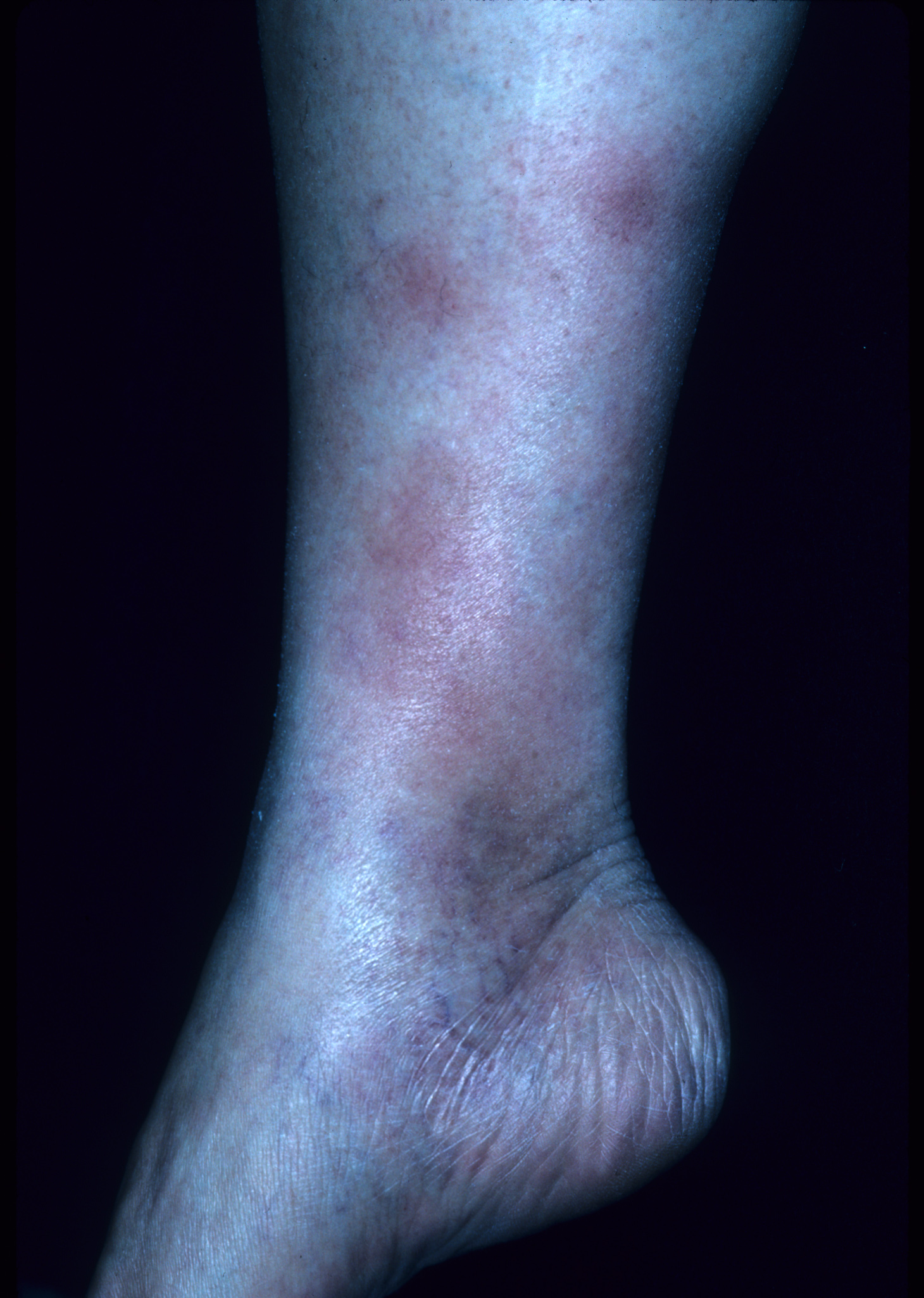
The patient’s rapid strep test was positive and she was diagnosed clinically with erythema nodosum (EN) secondary to group A beta-hemolytic streptococcal pharyngitis. EN is the most frequent type of septal panniculitis (inflammation of the septa of fat lobules in the subcutaneous tissue). EN tends to occur more often in women in the adult population, generally during the second and fourth decades of life.
While the exact percentage is unknown, one study estimated that 55% of EN is idiopathic. Identifiable causes can be infectious, reactive, pharmacologic, or neoplastic. Group A beta-hemolytic streptococcal pharyngitis has been linked to EN. A retrospective study of 129 cases of EN over several decades revealed that 28% of the patients had streptococcal infection. Nonstreptococcal upper respiratory tract infections may also play a role.
EN lesions are deep-seated nodules that may be more easily palpated than visualized. The lesions are initially firm, round or oval, and are poorly demarcated. As seen in this case, the lesions may be bright red, warm, and painful. The lesions number from 1 to more than 10 and vary in size from 1 to 15 cm. Lesions appear on the anterior/lateral aspect of both lower extremities. A characteristic of EN is the complete resolution of lesions with no ulceration or scarring. EN may occur with fever, malaise, and polyarthralgia.
The diagnosis of EN is mostly made on physical examination. When the diagnosis is uncertain, a biopsy that includes subcutaneous fat is performed. This can be a deep punch biopsy or a deep incisional biopsy sent for standard histology. For suspected strep cases, rapid strep test or throat cultures are best during acute illness, while antistreptolysin O titers may be used in the convalescent phase.
Look for, and treat, the underlying cause. There is limited evidence to guide treatment unless an underlying cause is found. In this case, the patient was treated with penicillin and nonsteroidal anti-inflammatory drugs and was advised to get some bedrest. She experienced complete resolution of the EN within 4 weeks.
Photos and text for Photo Rounds Friday courtesy of Richard P. Usatine, MD. This case was adapted from: Paulis R. Erythema nodosum. In: Usatine R, Smith M, Mayeaux EJ, et al., eds. The Color Atlas of Family Medicine. New York, NY: McGraw-Hill; 2009:756-759.
To learn more about The Color Atlas of Family Medicine, see:
* http://www.amazon.com/Color-Atlas-Family-Medicine/dp/0071474641